
Tag: ultrasound

Possibility of Early Spina Bifida Diagnosis from 10 Weeks.

Graphic of Early Spina Bifida Diagnosis. A baby that is getting its spine scanned at 10 weeks.

Spina Bifida Early Diagnosis from 10 Weeks of Pregnancy

DR Fred Ushakov Headshot. London Pregnancy Clinic Medical Director. Specialist in Fetal Medicine & Obstetrical Ultrasound.

An Interview with Dr. Fred Ushakov

Published

Tags

In honour of UK Spina Bifida Awareness Week, Dr. Ushakov, Founder and Managing Director of London Pregnancy Clinic, discusses the possibilities of early diagnosis of spina bifida. He is an internationally renowned expert in fetal medicine, with a primary focus on fetal spina bifida and is also currently affiliated with FMU UCLH. In recognition of UK Spina Bifida Awareness Week, we have had the privilege of asking Dr. Ushakov several vital questions about spina bifida.

Dr. Ushakov was interviewed by, our Specialist Sonographer in Fetal Medicine, Gynaecology & Fertility, Ms Shaz Khojasteh.

What sparked your interest in spina bifida?  

“In 1980, as a young obstetrician heavily involved in attending deliveries and particularly interested in ultrasound technology, I performed a scan on a young pregnant woman. I noticed something unusual about the baby’s brain, although I couldn’t quite discern the exact issue. Subsequently, I was present at her delivery, and to my surprise, the baby was born with spina bifida. This experience was truly shocking. It occurred over 35 years ago during the early stages of ultrasound technology when both knowledge and technology were limited.”

What actions did you take next?

“After that shocking experience, I became genuinely intrigued by effectively detecting spina bifida. I dedicated significant effort to mastering scanning techniques for identifying spina bifida in babies. Over time, I evolved into an expert in this field, with a particular focus on spotting it as early as 10 weeks into the pregnancy. And naturally, I made it a point to share this knowledge with my colleagues, advocating for early spina bifida detection.”

How many babies with spina bifida have you encountered?

“I’ve seen hundreds of fetuses with spina bifida during my career. It’s become a routine part of my work – conducting early scans to detect spina bifida and performing expert scans for babies already diagnosed with this condition. Spina bifida is actually relatively common. In the UK, it’s estimated that around 1 in every 1,000 pregnancies is affected by spina bifida. Even though it might not be as well-known as some other conditions, its effects on those who have it can be quite substantial. This is why early detection and awareness are so important.”

Why is spina bifida so important?

“Open spina bifida holds immense significance due to its prevalence as a major fetal structural defect. It adversely affects essential bodily functions, such as bowel and bladder control. Walking becomes a challenge for most babies born with open spina bifida, and some may also exhibit brain abnormalities. While treatments can enhance outcomes, a complete cure remains elusive. Many babies survive, but their lives are profoundly impacted by long-lasting disabilities. In essence, it’s a severe condition with enduring consequences.”

What do you consider the main issue with spina bifida?

“The primary concern regarding spina bifida, from my perspective, is the alarming lack of awareness. This lack of awareness extends not only to patients but also to primary healthcare providers. From my experience, many pregnant women take folic acid supplements without fully grasping their significance. It’s imperative to raise awareness and educate both the general public and healthcare professionals about this condition, its risk factors, and the crucial role of folic acid supplementation during pregnancy and early first-trimester spina bifida screening. Unlike the widely recognized Down’s syndrome, spina bifida remains relatively obscure. This is a significant concern because, for some babies with spina bifida, the potential outcomes can be even more challenging than those for Down’s syndrome.”

Fred, could you clarify why it’s essential to start taking folic acid immediately upon discovering pregnancy?

“Yes, immediate folic acid supplementation upon discovering pregnancy is of paramount importance. Neural tube formation is an early pregnancy event, usually concluding by the end of the 6th gestational week, approximately 28 days after conception. In babies with spina bifida, the neural tube fails to close correctly, leading to spinal cord and spine bone issues. This anomaly develops extremely early when the baby is less than 1 cm in size and has just initiated its heartbeat.

Folic acid plays a pivotal role during the critical period before and during the first six weeks of pregnancy when the baby’s spine is forming. In practical terms, most people discover their pregnancy around 4 weeks after conception, and by that point, they are already 4 weeks into the pregnancy. This leaves just two weeks during which folic acid can exert its most significant impact. Taking early action is the key to providing essential support for your baby’s neural tube development and minimizing the risk of spina bifida.”

Let’s delve into early Spina Bifida diagnosis during pregnancy. When can you identify this condition?

“With my level of expertise and the use of advanced 3D ultrasound scanners (Fig 1), I can diagnose the majority, probably over 90%, of open spina bifida as early as 10 weeks into the pregnancy. To put it differently, personally missing a diagnosis of spina bifida at that stage would be a great disappointment to me, and I would be highly motivated to conduct a thorough investigation into why such a diagnosis was missed. 

  • 3D Ultrasound GIF of baby at 10 week. Image by London Pregnancy Clinic - for educational purpose.

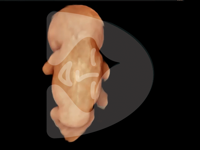
    Fig 1 – 3D ultrasound image of the baby at 10 weeks: All main structures are visible
  • It’s important to note that achieving a 100% accuracy rate in diagnosing spina bifida through ultrasound is a challenge. There can be significant variations in the presentation of spina bifida, and in some rare cases, the anomaly may go undiagnosed until delivery. While early detection is highly valuable, there are instances where the condition’s subtleties make it difficult to diagnose definitively with ultrasound.”

How can you diagnose spina bifida so early? Are you inspecting the baby’s spine?

“No, actually, it’s quite challenging to directly visualize spinal abnormalities at this early stage. What I’m primarily focused on is the fetal head and brain (Fig 2). In cases of open spina bifida, there’s a distinct phenomenon at play. There is a leakage of fluid from the brain through the hole in the spine, and this leakage creates a very specific brain anomaly known as Chiari 2 malformation. I’m essentially searching for the earliest evidence of this phenomenon in the ultrasound images. It’s a highly specific indicator – when this particular brain anomaly is present, it’s a strong indication that the baby has spina bifida.

Certainly, at 10 weeks, I also examine the fetal spine. The spine is not yet ossified, so it appears as dark lines running along the baby’s back, and often I can even discern individual vertebrae as bright dots (Fig 3). This level of detail means that very severe spinal deformities can be visible. Sometimes, spina bifida can manifest as a small bubble on the baby’s back at this early stage (Fig 4).”

  • Ultrasound GIF of baby's brain at 10 weeks. Image by London Pregnancy Clinic - for educational purpose.

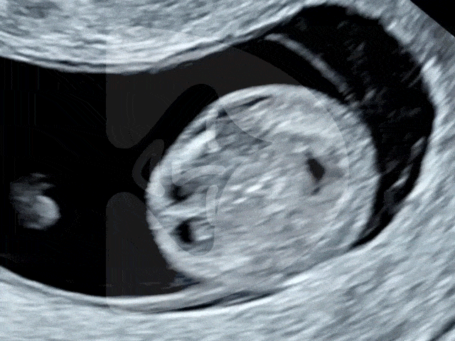
    Fig 2 – Normal brain at 10 weeks: this baby has no spina bifida
  • Ultrasound GIF of baby's spine at 10 weeks. Image by London Pregnancy Clinic - for educational purpose.

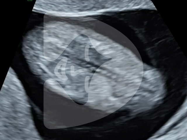
    Fig 3 – Baby’s spine at 10 weeks: no deformities
  • Ultrasound GIF of baby with spina bifida at 11 weeks. Spina bifida cystica is seen at the bottom of the spine. Image by London Pregnancy Clinic - for educational purpose.

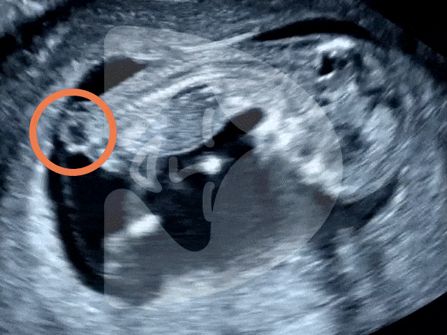
    Fig 4 – Baby with spina bifida at 10-11 weeks: spina bifida marked by the circle

Can other professionals detect spina bifida early?

“Yes, there are indeed many research studies indicating the possibility of early detection. One of the most significant research efforts comes from King’s College Hospital in London. Sonographers at this hospital have scanned over 100,000 babies at 11-13 weeks of pregnancy and were able to detect more than half of the fetuses with spina bifida. This achievement is truly impressive, although it’s important to note that it may not be applicable to all NHS trusts. King’s College Hospital has a dedicated unit, established protocols, and a comprehensive training program. Only a handful of other hospitals across the UK, including UCLH that I work for, have similar protocols in place.”

What is the situation like in an average NHS hospital?

“In NHS hospitals, there is a screening ultrasound program called the anomaly scan, which is typically conducted at 19-20 weeks into the pregnancy. During this scan, sonographers check for spina bifida among other anomalies.

Unfortunately, there isn’t a routine first-trimester screening for spina bifida, and the majority of cases are detected during the second trimester. In a 2019 survey of ultrasound units performing scans in the first trimester (11-13 weeks) in England, it was found that only 16% of them included a check of the fetal spine in their protocols.”

Tell us about the “crash sign” and its significance.

“Yes, indeed. Many research groups have proposed various methods for detecting spina bifida in the first trimester. In our case, we’ve come up with a marker that represents the ultrasound appearance of Chiari 2 malformation in the early fetal brain. It’s pretty obvious when you have a top-notch ultrasound scanner.

Now, why ‘CRASH’? We actually have a video up on YouTube that demonstrates it, and once you watch it, the name will make perfect sense.”

Any advice for patients seeking early spina bifida ruling out?

“If you’re keen to know early on, I’d suggest considering booking a 10 Week Scan with a professional having a special interest in this area. For those with previous pregnancies affected by spina bifida, arranging a 10 Week Scan at Fetal Medicine Unit at UCLH with a referral from your primary NHS care provider is an excellent choice. Alternatively, you can book a 10 Week Scan privately at London Pregnancy Clinic. At our clinic, we can combine this scan with non-invasive prenatal testing (NIPT). Please inform our admin staff about your worries, and we will arrange a proper scan for your baby.

If you prefer to stick with NHS services, ask your sonographer during the 11-13 weeks scan if they’re actively screening for spina bifida. Some hospitals in London may have established protocols for this. However, if your NHS provider doesn’t routinely check for spina bifida (which is the case for most hospitals), you might want to consider an early anomaly scan (Early Fetal Scan) at 13-16 weeks through a private provider. For those going the private scan route, it’s a good idea to check if the clinic screens for spina bifida at early stages. Advanced private ultrasound clinics are typically well-equipped, and their staff may have special training in fetal medicine.

Please note that the majority of sonographer-run clinics that provide NIPT or perform non-medical gender scans do not screen for spina bifida.”

It seems like you prefer screening for spina bifida at 10 weeks rather than at the time of the standard NHS scan around 12 weeks. Is that accurate, and could you tell us why?

“Yes, you’ve got it right. In the past, I used to advocate for spina bifida screening during the conventional nuchal translucency scan at around 12-13 weeks. But with the introduction of NIPT, which can be done as early as 10 weeks, I’ve started scanning for spina bifida even before the blood test. At 10 weeks, the little ones are really tiny, measuring less than 1.5 inches (around 35 mm).

Interestingly, with the help of state-of-the-art ultrasound technology (and we’re lucky to use the most advanced machines), many fetal structures are surprisingly easier to examine at 10 weeks compared to 12 weeks. The brain and spine, in particular, stand out as more visible structures.

I won’t bore you with all the technical stuff, but it’s somewhat paradoxical that I feel that an early screening at 10 weeks is actually probably more effective. Plus, it’s a relief for parents who are understandably anxious and want reassurance as soon as possible. But it’s essential to recognize that this type of screening demands a high level of expertise and top-notch ultrasound technology to be done effectively.”

Can other professionals detect spina bifida early?

“Yes, indeed. There are internationally renowned groups in Melbourne, Nice, and Berlin, among others, who also have experience in diagnosing spina bifida before the 11th week of pregnancy. At present, our joined expertise is largely based on anecdotal cases. This is because the incidence of spina bifida is relatively rare, occurring at a rate of 1 in 1,000 pregnancies. To establish statistically significant numbers and determine the detection rate of a 10-week scan, one would need to scan thousands and thousands of babies.”

What is the SMART TEST, and does it include screening for spina bifida?

“The SMART TEST is an innovative concept in prenatal care developed and offered by the London Pregnancy Clinic. It combines extended NIPT with expert ultrasound. The SMART TEST is unique in that it covers a wide range of structural anomalies, chromosomal conditions, and genetic diseases, and it can do so as early as 10 weeks into the pregnancy.

A primary target of the SMART TEST is indeed the earliest possible screening for spina bifida. The test incorporates advanced 2D and 3D ultrasound technology to thoroughly examine the fetal brain and spine at a stage when the baby has just transitioned from an embryo to a fetus. This comprehensive approach ensures early detection and peace of mind for expectant parents.”

Why do you believe that early detection of spina bifida is so crucial? After all, the condition can be diagnosed by the NHS anomaly scan at 20 weeks.

“I’m a strong advocate for early detection of spina bifida, and let me explain why. Spina bifida is a highly serious structural anomaly associated with severe disabilities. While we’ve made significant progress with in-utero surgical treatments, it’s important to understand that these treatments don’t cure the condition.

Moreover, it’s essential to highlight the significance of the surgical aspect. At UCLH, I’m part of the Fetal Surgery for Spina Bifida team, which is led by Professor Jan Deprest, an internationally renowned expert in fetal surgery. Our centre is the sole one in the UK commissioned by the NHS to provide this cutting-edge treatment. In my role, I conduct scans on the baby before and after the surgery. I take great care to evaluate the spinal lesion, perform a thorough examination of the baby’s brain, and perform a comprehensive top-to-toe scan of the entire baby. I’ve had the privilege of scanning hundreds of babies with spina bifida before and after surgery, contributing to their care.

Performing surgery on a baby with spina bifida while they’re still in the mother’s womb is an incredible feat. This complex procedure requires a skilled international team of doctors and medical professionals from the UK and Belgium. The delicate balance of providing life-changing treatment for the baby while ensuring the safety of both the baby and the mother is a remarkable achievement in fetal medicine.

However, almost half of mothers are not eligible for this ground-breaking surgical treatment, often due to the severity of the baby’s condition or additional fetal problems. For these parents, making difficult decisions about the pregnancy becomes a reality, often after 20 weeks. Even for parents eligible for surgery, the journey is challenging. They go through numerous tests, counselling, and procedures in a short time, adding to the stress.

All of this can be alleviated with early diagnosis. Early detection provides parents with the time and information they need to make informed decisions. For the most severe cases of spina bifida that are not operable, early detection helps parents understand their options from the start of the pregnancy. That’s why I advocate for screening for spina bifida as early as 10 weeks or at least at 12 weeks.”

Fred, as a conclusion, what advice or suggestions would you like to offer to future parents?

“If you are in the planning stages of pregnancy, consider starting to take folic acid supplements. Folic acid is most effective when taken before conception and during the first few weeks of pregnancy when the baby’s spine is forming. This simple step can contribute to the healthy development of your baby and reduce the risk of spina bifida.

Additionally, I’d like to emphasize the importance of early screening for spina bifida, which can now be done as early as 10 weeks into the pregnancy. This can provide you with valuable information, peace of mind, and ample time to make informed decisions about your baby’s health. If you have concerns or a history of spina bifida in your family, consider the SMART TEST at the London Pregnancy Clinic. It’s a comprehensive approach that combines NIPT and advanced ultrasound technology to screen for a wide range of conditions, including spina bifida.

Remember that knowledge is power. Being informed and seeking early detection can make a significant difference in the journey of your pregnancy. Stay well-informed, ask questions, and don’t hesitate to seek expert guidance. Your baby’s health is our priority, and we are here to help you every step of the way.”

References

(1) Karim, J., Pandya, P., McHugh, A. and Papageorghiou, A.T. (2019). OC23.06: Significant variation in practice for first trimester anatomy assessment: results from a nationwide survey. Ultrasound in Obstetrics & Gynecology, 54(S1), pp.60–61.

(2) Ushakov, F., Sacco, A., Andreeva, E., Tudorache, S., Everett, T., David, A.L. and Pandya, P.P. (2019). Crash sign: new first-trimester sonographic marker of spina bifida. Ultrasound in obstetrics & gynecology : the official journal of the International Society of Ultrasound in Obstetrics and Gynecology, [online] 54(6), pp.740–745.

(3) University College London Hospitals NHS Foundation Trust. (n.d.). Management of Fetal Spina Bifida. [online] Available at: https://www.uclh.nhs.uk/patients-and-visitors/patient-information-pages/management-fetal-spina-bifida [Accessed 21 Oct. 2023].

(4) Syngelaki, A., Hammami, A., Bower, S., Zidere, V., Akolekar, R. and Nicolaides, K.H. (2019). Diagnosis of fetal non‐chromosomal abnormalities on routine ultrasound examination at 11–13 weeks’ gestation. Ultrasound in Obstetrics & Gynecology, 54(4), pp.468–476.

(5) Rolnik, D.L., Wertaschnigg, D., Benoit, B. and Meagher, S. (2020). Sonographic detection of fetal abnormalities before 11 weeks of gestation. Ultrasound in Obstetrics & Gynecology, 55(5), pp.565–574.

Latest Stories

Continue reading

Spina Bifida Awareness Week

Graphic of Spina Bifida Awareness Week. A baby that is getting its spine scanned at 10 weeks.

Spina Bifida Awareness Week:

What is it? and how we can screen for it?

Published

Tags

This week is Spina Bifida Awareness Week, a time to shine a light on this important condition. We look at what Spina Bifida is, how it can be detected and charities that spread awareness about it.

Comprehensive Screening:  

From the 16th to the 22nd of October, we shine a light on Spina Bifida Awareness Week. But first, let’s break it down. What exactly is spina bifida? It’s a condition where the spine doesn’t fully develop in the womb. It occurs when the spine and spinal cord fail to develop properly in the womb, resulting in a gap in the spine. This incomplete development can lead to both physical and intellectual challenges.

A surprising fact? As many as 10% of people might have a milder form of spina bifida. Most aren’t even aware of it. On the more severe end of the spectrum, open spina bifida impacts about 700 babies in the UK every year.

Now, here’s the uplifting part. Taking folic acid during the early stages of pregnancy can significantly reduce the risk of a baby developing spina bifida. However, many women still don’t know about this simple preventative measure. It’s essential for healthcare providers to share this knowledge.

Early Detection Makes a Difference! 

At our clinic, we’re proud to offer a specialised Ten-week Anomaly Scan. This scan is designed to detect signs of spina bifida at a very early stage. By identifying it early, families can make informed decisions and medical professionals can provide the best care options. Early detection can dramatically improve outcomes for affected pregnancies. Surgeons from UCLH and GOSH carried out the first procedure on babies in the womb in the UK back in 2018. This international team received their training at UZ Leuven1

While many might be more familiar with conditions like Down Syndrome, it’s vital to bring spina bifida to the forefront as well. Its prevalence is notable, and with the right measures like early detection and folic acid intake, we can make a difference.

Join Our Mission in Boosting Awareness: 

Knowledge is power. By understanding spina bifida, its risks, and the preventive steps, we can safeguard future generations. Let’s raise the bar of awareness together by educating ourselves and supporting charities like Shine. 

Shine Charity is a UK-based charity that supports people with spina bifida and hydrocephalus. The charity’s vision is a society where all those affected by spina bifida and/or hydrocephalus are empowered and enabled. Shine’s mission is to make a positive difference to the lives of people affected by these conditions.

Shine’s goals for 2022-2027 include:

  • Enabling babies and children with spina bifida and/or hydrocephalus to achieve their potential
  • Empowering young people with these conditions
  • Ensuring that adults with these conditions have the choices and opportunities to live healthy and independent lives

Watch the explainer video on the 10 week scan:

Latest Stories

Continue reading

NIPT and Scan: Why We Champion This Dual Approach

Graphic of a baby footprint in a heart with DNA strand on either side.

NIPT and Scan Approach: Why We Champion This Screening Method at London Pregnancy Clinic

Published

Tags

At the London Pregnancy Clinic, we’re dedicated to providing expectant mothers with advanced screening options and the highest level of care. We firmly recommend combining Non-Invasive Prenatal Testing (NIPT) and Ultrasound screening. Let’s explore why we endorse NIPT and Scan approach and how it benefits our patients.

Why Choose Both NIPT and scan?

Comprehensive Screening:  

Ultrasound visually assesses the baby’s anatomy, checking for physical abnormalities and measuring growth. NIPT, known by brand names like Natera’s Panorama AI or Eurofins’ PrenatalSafe, examines fetal DNA in the mother’s bloodstream, providing insights into potential chromosomal abnormalities like Down’s Syndrome, Edwards syndrome, and Patau syndrome.

Increased Accuracy and Early Detection:  

By merging Ultrasound’s structural insights with genetic data from NIPT, we significantly reduce false positives and offer more accurate results. As early as 10 weeks, when your baby is the size of a strawberry, we initiate the dual screening process. At this stage, we conduct the earliest possible structural anomaly scan, the Ten-week Anomaly Scan, to search for structural anomalies that NIPT can’t detect. We can rule out severe physical abnormalities like Acrania, Spina bifida, Absence of arms, hands, legs or feet, and Alobar holoprosencephaly. Only after confirming your baby’s structural development do we proceed with the NIPT test.

UNDERSTANDING THE TECHNOLOGY

Ultrasound Screening: 

Ultrasound employs sound waves to create images of the baby in the womb. A small probe, called a transducer, moves over the mother’s abdomen. The transducer emits high-frequency sound waves that bounce off the baby’s structures, and these echoes are converted into images on a screen.

Non-Invasive Prenatal Testing (NIPT): 

NIPT is a simple blood test taken from the expectant mother. This test detects tiny fragments of the baby’s DNA circulating in the mother’s bloodstream. By analysing these fragments, we can determine the risk of certain chromosomal conditions.

Is It Safe?

Absolutely. Both Ultrasound and NIPT are non-invasive and pose minimal to no risk to both mother and baby. However, it’s important to note that while NIPT is highly effective, it’s not a definitive diagnostic test. In cases of low negative predictive value, our doctors may recommend invasive tests like CVS or amniocentesis, which carry minimal miscarriage risk.

Our NIPT Options

As early as…
  • 10 weeks

  • 9 weeks

  • 10 weeks

Turnaround (Working Days)
  • 2-4

  • 5-7

  • 5-7

Lab Location
  • UK

  • US

  • US

No Call Results
  • <1%

  • <1%

  • <1%

Redraw Rate
  • 2%

  • 3%

  • 2%

Edward’, Patau & Down’s Syndrome
Di George Syndrome (22q del)
Triploidy
Turner Syndrome (45X)
Sex chromosomes aneuploidies
Twin pregnancies
  • Best

Vanishing twin syndrome
Fetal sex reveal (optional)
Scan + NIPT Price
  • £540

  • £540

  • £490

Extended NIPT + Scan Options
  • SMART Test £1690

  • Microdeletions £790

  • Rare Diseases £790

Other Early Ultrasound Screenings Offered

For those looking to delay their first scan, London Pregnancy Clinic offers pioneering Early Ultrasound Screenings, including the Early Fetal Scan conducted between 12 and 16 weeks, which can exclude more than one hundred serious anomalies. Additionally, our Early Fetal Echocardiography is designed to identify up to 80% of detectable severe fetal heart defects. It is a scan we highly recommend this scan for all babies with increased nuchal translucency (NT) measurements, fetal anomalies, or other unusual findings detected at 11-13 weeks scan.

Conclusion

At the London Pregnancy Clinic, we believe in providing the most comprehensive care possible. By endorsing the dual Ultrasound and NIPT approach, we ensure that our patients receive a detailed, accurate, and safe assessment of their baby’s health. Whether you choose the ten-week scan or another early anomaly scan, we’re here to guide and support you every step of the way.

If you have further questions or would like to schedule an NIPT and scan, please contact the London Pregnancy Clinic.

Book NIPT + Scan from £490

Latest Stories

Continue reading

Harmony NIPT Test – Yesterday’s News

Harmony NIPT Test – Yesterday’s News

Published

Tags

TDL Genetics ends Harmony NIPT provision after a decade. With outdated technology, high no-call rates, and fierce competition, Harmony loses its shine. As leading NIPT providers, we’re reviewing TDL’s new offering. But for now, our advanced menu offers the latest prenatal screening. Remember – ultrasound still crucial for detecting 2/3 of fetal abnormalities.

End of an era…

TDL Genetics (The Doctor’s Laboratory) have announced last week that it will cease providing Harmony NIPT in its UK laboratories. TDL started providing Harmony Non-Invasive Prenatal Screening back in 2013.

Harmony NIPT, which was introduced by Ariosa Diagnostics in 2013, was for a long time a gold standard in Non-Invasive Prenatal Testing. The company was later acquired by the Swiss giant Roche Holdings, which was very successful at marketing the test and in many countries, the brand name ‘Harmony Test’ has become synonymous with ‘NIPT’. In the UK, this was particularly the case as it was one of the first NIPT tests to be approved by the NHS, and it offered in many state hospitals.

NIPT was first introduced by the now-defunct Sequenom, using the brand name MaterniT21Plus™, which was approved by the FDA in 2011. Harmony Test pioneered a new method of NIPT screening using a different cfDNA sequencing method. Harmony NIPT exhibited excellent clinical performance data at the time, helping it solidify its status as the preferred NIPT test by many healthcare professionals.

The future looks good!

In the past 10 years, NIPT has become a heavily researched space, meaning that many companies have raced to create their own NIPT test using a host of advanced technologies. A couple of such competitors are Natera’s ‘Panorama Test’ and Erofins’ ‘PrenatalSAFE Test’. This increased level of competition meant that patients could now benefit from much more advanced tests for chromosomal anomalies such as Down’s syndrome and screening for rare genetic diseases.

In this time, Harmony NIPT has fallen behind in terms of its technology versus the new tests. Furthermore, the equipment used for the test has become dated and as such many of the tests performed by TDL ended up failing to produce conclusive results due to ‘quality control issues’. These so-called ‘no-call’ results, or failure of one of the testing tubes, have become so prevalent with the samples we sent to TDL that we raised our first of many complaints with TDL back in 2021. Unfortunately, these lab challenges caused significant emotional distress for our expectant parents and frustration for our clinic’s team.

NIPT at London Pregnancy Clinic

In light of these developments, at London Pregnancy Clinic, being one of the top NIPT providers in the UK and leading international experts in fetal medicine, we set on the path of finding the best alternative NIPT. We were one of the first clinics in the UK to offer the advanced NIPT test from Invitae back in 2021. Since then, we have worked with many NIPT brands and have helped hundreds of patients navigate the best NIPT choice for them.

Our clinical team has spent months interviewing the laboratory teams of all major NIPT providers as well as auditing their clinical performance. We are now the leading provider of NIPT in London, and we feel responsible for our patients’ pathway to choose the best option in the market at every given time. We have the experience of providing the Harmony NIPT and other NIPTs, and the expertise to distinguish the performance of each test in the market. We do not offer any NIPT without thoroughly reviewing its characteristics and clinical data.

As of TDL’s new NIPT option, namely the Illumina VeriSeq NIPT, we are still waiting for the lab to send us clinical performance data and set up an interview with the lab team before we are able to offer the test to our patients. For now, we have many questions for the lab given the poor quality of performance of the Harmony NIPT in terms of no-call results which was much higher than Roche’s advertised levels from our experience. In the meantime, we are confident that our current menu of NIPT tests is the latest and most advanced screening options in the market with similar turnaround times to TDL.

As always, we will keep reminding our patients that NIPT should be done alongside a complete examination of the fetal anatomy via an ultrasound scan. The NIPT marketing machine has helped raise awareness of certain chromosomal anomalies, in particular Down’s Syndrome. However, NIPT can give you a false sense of reassurance as it is useless in screening for structural anomalies (physical defects) representing more than 2/3 of known fetal abnormalities and can often be detected using ultrasound.

Latest Stories

Continue reading

A Journey Through the First 10 Weeks of Pregnancy: Week-by-Week Guide & Ultrasound Scans

The first 10 weeks of pregnancy are truly transformative. As the foetus develops rapidly during this period, the significance of each week becomes ever more pronounced. If you’re an expectant mother or just curious about this marvellous journey of life, join us as we navigate through the initial 10 weeks, emphasising the value of ultrasound pregnancy scans.

Week 1 & 2: Although these weeks precede conception, they mark the beginning of the pregnancy timeline, starting from the first day of your last menstrual cycle.

Ultrasound Scan: Generally, no scans take place during these weeks. However, it’s an opportune moment to see your GP and plan your pregnancy journey. Consider Beta HCG pregnancy test.

Week 3: The magic begins! The sperm meets the egg, resulting in a zygote, which embarks on its journey through the fallopian tube.

Ultrasound Scan: It’s early days, and while an ultrasound won’t show much, an at-home test could provide the first inklings of pregnancy.

Week 4: As the zygote morphs into a blastocyst and finds its home in the uterus, the baby’s development begins in earnest.

Size: Not larger than a poppy seed.

Ultrasound Scan: Some women may opt for an early transvaginal scan which might show the start of a gestational sac.

Week 5: Layer separation occurs in the embryo, signalling the beginning of the baby’s organ development. The neural tube, the precursor to the spine and brain, begins to form.

Size: Tiny, approximately the size of a sesame seed.

Growth Factor: Grown about 2 times from the previous week.

Ultrasound Scan: A transvaginal scan can now possibly show the gestational sac. If you’re exhibiting symptoms of ectopic pregnancy, consider a scan (pain low down and on one side of your abdomen, bleeding from your vagina, pain in the tip of your shoulder, discomfort when going to the toilet, a brown watery discharge from your vagina.)

Week 6: With the formation of limb buds and a heart that’s started beating, this week is crucial.

Size: As large as a lentil.

Growth Factor: Grown about 3 times from the previous week.

Ultrasound Scan: A heartwarming moment for many, as the baby’s heartbeat may be visible! Consider our Viability Scan.

Week 7: Brain development accelerates and facial features start shaping up.

Size: About the size of a blueberry.

Growth Factor: Grown about 2 times from the previous week.

Ultrasound Scan: The scan reveals an increasingly defined shape, including a clearer heartbeat. Book our Viability scan from this point to 9 weeks.

Week 8: The foetus now has tiny fingers and toes, and the skeletal structure begins to form.

Size: Almost as big as a raspberry.

Growth Factor: Grown by 1.5 times from the previous week.

Ultrasound Scan: Budding limbs are now visible, making this scan a particularly exciting one.

Week 9: Most of the baby’s critical body parts are now in place and will keep maturing.

Size: Comparable to a cherry.

Growth Factor: Grown about 1.3 times from the previous week.

Ultrasound Scan: The baby’s shape and movements become more evident, although they can’t be felt just yet.

Week 10: Muscular and skeletal structures continue to solidify, and the tiny nails begin to emerge.

Size: Almost the size of a strawberry.

Growth Factor: Grown by 1.2 times from the previous week.

Ultrasound Scan: Now, you can witness finer details of your baby’s development, from the nails to its more discernible body shape. Book our 10 week scan, and consider NIPT.

To sum up: From a microscopic beginning to the size of a strawberry, the journey of the first 10 weeks of pregnancy is nothing short of miraculous. Ultrasound scans become a window to this evolving world, illustrating the baby’s swift progress. As the journey continues, prioritising health and staying informed during these foundational weeks is of paramount importance. The adventure has only just commenced!

Gynaecological Pelvic Scans: Importance, Procedure, and Diagnostics

In the realm of gynaecology, pelvic scans play a crucial role in maintaining women’s health. These essential imaging tests help visualise the organs within the female pelvic region, aiding the diagnosis of various conditions. This comprehensive blog post provides an in-depth understanding of gynaecological pelvic scans – their importance, procedure, potential risks, the conditions they can diagnose, their history, and the organs they help visualise.

Why Are Gynaecological Pelvic Scans Important?

Gynaecological pelvic scans provide detailed images of the female pelvic organs, including the uterus, ovaries, fallopian tubes, cervix, and bladder. This non-invasive procedure aids in the diagnosis and management of numerous conditions such as fibroids, ovarian cysts, endometriosis, pelvic inflammatory disease (PID), and even cancer.

This type of imaging allows medical professionals to identify abnormalities, track their progress, and evaluate the effectiveness of treatments. It’s also a vital tool for pregnancy management, helping to monitor foetal development, placental health, and diagnose ectopic pregnancies.

How are Gynaecological Pelvic Scans Performed?

Pelvic scans in gynaecology are typically performed through ultrasound technology. Ultrasounds work by emitting high-frequency sound waves that bounce off tissues and organs, creating echoes that are converted into real-time images.

There are two main types of gynaecological pelvic scans: transabdominal and transvaginal. The transabdominal scan is performed externally, with a probe moved over the lower abdomen. For a transvaginal scan, a specially designed probe is inserted into the vagina, providing closer, more detailed images of the pelvic organs.

Are There Any Risks? Are They Painful?

Gynaecological pelvic scans are generally safe, non-invasive procedures with minimal risk. Ultrasound technology does not involve radiation, making it safer than other imaging techniques.

In terms of discomfort, while some women might experience mild discomfort during a transvaginal scan, it’s typically not painful. The procedure is usually quick, and any discomfort tends to subside immediately after the scan.

Is There a Need for Preparation?

Preparation for a gynaecological pelvic scan may vary depending on the type of ultrasound. For a transabdominal scan, patients may be asked to drink water before the procedure to fill the bladder, which allows better visualisation of the pelvic organs. For a transvaginal scan, typically, no specific preparation is required.

Conditions Diagnosed by Gynaecological Pelvic Scans

Pelvic scans can diagnose a multitude of conditions, including:

  1. Fibroids: Non-cancerous growths in the uterus, often appearing during childbearing years.
  2. Ovarian cysts: Fluid-filled sacs in the ovary, usually harmless but occasionally requiring treatment.
  3. Endometriosis: A condition where tissue similar to the lining of the uterus grows outside it, causing pain and potentially affecting fertility.
  4. Pelvic Inflammatory Disease (PID): An infection of the female reproductive organs, often due to sexually transmitted bacteria.
  5. Gynaecological cancers: Pelvic scans help detect and manage cancers of the cervix, uterus, and ovaries.

The History of Scans in Gynaecology

The use of ultrasound technology in gynaecology has evolved significantly since its first clinical application in the 1950s. The initial ‘A-mode’ ultrasounds, which provided one-dimensional information, evolved into ‘B-mode’ in the late 1950s, delivering two-dimensional images. The introduction of Doppler ultrasound in the 1970s brought colour to the images, providing information about blood flow. Today, we even have 3D and 4D ultrasounds, allowing for detailed three-dimensional imaging and real-time movement.

Organs Visualised by Gynaecological Pelvic Scans

Gynaecological pelvic scans offer comprehensive imaging of the female pelvic organs. This includes the uterus, ovaries, fallopian tubes, cervix, and bladder. Another type of scan that specifically focuses on the potency of the fallopian tubes is know as HyCoSy.

Unravelling Microdeletions: Their Causes, Risk Factors, Common Types, and Screening Methods

Microdeletions are a topic of extensive genetic research. These small deletions in the DNA sequence of a chromosome can have significant implications on an individual’s health. In this comprehensive blog post, we delve into the causes, risk factors, ten most common types of microdeletions, their prevalence, and the role of Non-Invasive Prenatal Testing (NIPT) in their detection.

What are Microdeletions?

Microdeletions are tiny losses of genetic material in the DNA sequence of a chromosome. These minute changes are typically undetectable under a standard microscope, hence the term ‘micro’. However, despite being minute, these deletions can lead to significant health and developmental problems, as they may disrupt several genes essential for growth and development.

Causes and Risk Factors

The exact cause of microdeletions remains unknown, and their occurrence seems largely spontaneous. They typically occur during the formation of reproductive cells or in early foetal development, resulting from a mistake in the cell’s DNA replication process.

In terms of risk factors, there’s no established link between microdeletions and parental age, ethnicity, or lifestyle. These changes can happen in any pregnancy, but familial history can increase the chances if a parent has a balanced translocation or an inherited microdeletion.

The Ten Most Common Microdeletion Syndromes

Here’s a list of ten of the most common microdeletion syndromes, along with their estimated prevalence:

SyndromeApproximate Prevalence
1. DiGeorge Syndrome (22q11.2 deletion syndrome)1 in 3,000 – 1 in 6,000
2. Williams Syndrome (7q11.23 deletion syndrome)1 in 7,500 – 1 in 20,000
3. Prader-Willi Syndrome1 in 10,000 – 1 in 25,000
4. Angelman Syndrome1 in 10,000 – 1 in 20,000
5. Cri-du-chat Syndrome (5p- syndrome)1 in 20,000 – 1 in 50,000
6. 1p36 Deletion Syndrome1 in 5,000 – 1 in 10,000
7. Wolf-Hirschhorn Syndrome (4p- syndrome)1 in 20,000 – 1 in 50,000
8. Smith-Magenis Syndrome (17p11.2 deletion syndrome)1 in 15,000 – 1 in 25,000
9. Alagille Syndrome (20p12 deletion syndrome)1 in 30,000 – 1 in 50,000
10. Rubinstein-Taybi Syndrome1 in 100,000 – 1 in 125,000

Microdeletions, in general, occur in approximately 1 in every 1,000 births, making them a significant contributor to genetic disorders. Just to compare Down’s Syndrome, the most common chromosomal disorder affects approximately 1 in 1,000 to 1 in 1,100 live births worldwide. The frequency of Down’s Syndrome increases significantly with the mother’s age, particularly from 35 years onwards whereas microdeletions are believed to occur randomly.

Microdeletion Screening: The Role of NIPT

Screening for microdeletions has been revolutionised by advancements in genetic testing, particularly with the introduction of Non-Invasive Prenatal Testing (NIPT). This screening test, performed from the 10th week of pregnancy, analyses cell-free DNA in the maternal blood to identify common chromosomal abnormalities and microdeletions. NIPT poses no risk to the foetus and offers a safer alternative to invasive diagnostic procedures.

Though NIPT has high accuracy rates, it’s crucial to understand that a ‘positive’ result indicates an increased risk, not a definitive diagnosis. Positive NIPT results should always be confirmed through diagnostic tests such as amniocentesis or chorionic villus sampling (CVS).

There are a number of NIPT providers who offer screening for microdeletions, such as Panorama NIPT, Unity NIPT and PrenatalSAFE.

Conclusion

While the world of microdeletions might seem complex, understanding them is crucial to advancing our knowledge of genetic disorders. Early detection, made possible by advanced screening methods like NIPT, allows for better preparation and potential intervention. As research continues to evolve, we can hope for even more effective detection and management of these genetic changes, ultimately leading to improved healthcare and patient outcomes.

Remember, if you have any concerns or questions about genetic disorders or prenatal testing, it’s always advisable to consult with a healthcare professional who can provide tailored advice based on your individual circumstances.

DiGeorge Syndrome: Causes, Risk Factors, Screening and Prevalence

DiGeorge Syndrome, also commonly known as 22q11.2 deletion syndrome (or simply 22q del,) is a complex and multifaceted disorder that many people may not be aware of. This blog post aims to shed light on the causes and risk factors of DiGeorge Syndrome, discuss screening options such as Non-Invasive Prenatal Testing (NIPT), and compare its prevalence with conditions such as Down’s Syndrome.

Understanding DiGeorge Syndrome (22Q DEL)

DiGeorge Syndrome is a chromosomal disorder caused by the deletion of a small piece of chromosome 22, specifically on the q11.2 region. The deletion happens spontaneously during the formation of reproductive cells or in early fetal development. The primary cause of 22q del is unknown, and it typically isn’t inherited from the parents.

The syndrome is characterised by a wide range of potential symptoms, including heart defects, certain facial features, and learning difficulties.problems with immune system and other abnormalities. Unfortunately, 22q del syndrome is also associated with learning difficulties and psychiatric or behavioural problems like autism and schizophrenia. Due to its complex nature, different individuals affected may present a different set of symptoms, making it a highly variable condition.

Risk Factors

Although the exact cause of the chromosomal deletion leading to DiGeorge Syndrome is unknown, it’s not typically associated with the age of the parent, unlike some other genetic disorders. The occurrence appears to be mostly random, which means that all pregnancies, irrespective of familial history, have a minimal but real risk.

Screening for DiGeorge Syndrome: The Role of NIPT

Non-Invasive Prenatal Testing (NIPT) has emerged as an invaluable tool for the early detection of several genetic disorders, including DiGeorge Syndrome. NIPT analyses cell-free DNA in the maternal blood to detect common chromosomal abnormalities. This test can be performed from the 10th week of pregnancy and poses no risk to the fetus.

While NIPT is highly accurate for detecting common trisomies like Down’s Syndrome (trisomy 21), Edwards’ syndrome (trisomy 18), and Patau’s syndrome (trisomy 13), it’s also valuable for identifying deletions like those causing DiGeorge Syndrome. It’s essential to remember, however, that a positive NIPT result for DiGeorge syndrome should be confirmed with diagnostic tests like amniocentesis or chorionic villus sampling (CVS) for a definitive diagnosis.

Not all NIPT provider offer reliable screening for DiGeorge Syndrome. Recently, the Panorama AI NIPT demonstrated impressive clinical performance for screening Di George Syndrome, you can read more about the study here. In well-designed prospective study Panorama Test was able to detect more than 80% of the fetuses affected by 22q del, showing an outstanding 50% positive predicted value.

Prevalence of DiGeorge Syndrome vs Down’s Syndrome

DiGeorge Syndrome is considered one of the most common genetic syndromes, second only to Down’s Syndrome. In the general population, it’s estimated to affect between 1 in 2,000 to 1 in 4,000 live births. Younger women have the same chance to deliver baby with 22q del as older ones.

In contrast, Down’s Syndrome, the most common chromosomal disorder, has a higher prevalence rate, affecting approximately 1 in 1,000 to 1 in 1,100 live births worldwide. The frequency of Down’s Syndrome increases significantly with the mother’s age, particularly from 35 years onwards.

Conclusion

Understanding and awareness of genetic disorders like DiGeorge Syndrome are crucial, not just for healthcare professionals, but for the general public too. While DiGeorge Syndrome is less common than Down’s Syndrome, it still represents a significant proportion of genetic disorders. By employing advanced screening methods such as NIPT, early detection and management of these conditions can be made possible.

Remember, if you have concerns or questions about genetic disorders or prenatal testing, it’s always best to consult with a healthcare professional who can provide advice tailored to your individual circumstances.

Understanding The Statistics Behind NIPT (Non-Invasive Prenatal Testing)

Summary:

The best NIPT will be really good at detecting problems (high sensitivity) and correctly saying everything is fine (high specificity). It should hardly ever make mistakes by saying there’s a problem when there isn’t (low false positive) or missing a problem and saying everything is okay (low false negative). If the test says there’s an issue, it should be highly likely to be true (high positive predictive value). And if it says everything is fine, it should be trustworthy (high negative predictive value).

NIPT (non-invasive prenatal testing) is a type of genetic test used to screen for certain chromosomal abnormalities and genetic syndromes in a developing fetus. The test involves analyzing a sample of the mother’s blood to look for fragments of DNA from the fetus.

It’s good to know the statistical terms related to NIPT performance because they help you understand how accurate the test is and what the results mean. You might be wondering why it’s important to understand those “boring statistics” related to NIPT (non-invasive prenatal testing) performance, but trust us, it’s actually really helpful! Knowing these statistical terms can make a big difference in your pregnancy journey.

By understanding these terms, you can make informed choices about your prenatal care. If the test is really accurate, you might feel more confident in the results and decide not to have further tests. But if the test has limitations, you might want to consider additional testing to get more clarity. It’s all about making sure you have the information you need to make the best choices for you and your baby.

So, while statistics might seem boring at first, they play a vital role in your understanding of the test’s accuracy, interpretation of results, and decision-making process. Embracing these statistics can give you more confidence and peace of mind throughout your pregnancy journey.

Please see below the most important statistical terms related to NIPT performance:

  1. Sensitivity: This measures how good the test is at correctly identifying fetuses that have a chromosomal abnormality. A high sensitivity means that the test is good at detecting most cases of abnormality. For example, if a test has a sensitivity of 99%, it means that out of 100 fetuses with a chromosomal abnormality, the test correctly identifies 99 of them.
  2. Specificity: This measures how good the test is at correctly identifying fetuses that do not have a chromosomal abnormality. A high specificity means that the test avoids identifying normal fetuses as abnormal. For example, if a test has a specificity of 99%, it means that out of 100 normal fetuses, the test correctly identifies 99 of them as normal.
  3. Positive predictive value (PPV): This is the probability that a positive test result is a true positive. In other words, if the test comes back positive, the PPV measures the likelihood that the fetus actually has a chromosomal abnormality. For example, if a test has a PPV of 95%, it means that out of 100 positive test results, 95 of them are true positives.
  4. Negative predictive value (NPV): This is the probability that a negative test result is a true negative. In other words, if the test comes back negative, the NPV measures the likelihood that the fetus does not have a chromosomal abnormality. For example, if a test has an NPV of 99%, it means that out of 100 negative test results, 99 of them are true negatives.
  5. False positive rate (FPR): This measures how often the test gives a positive result for a normal fetus. A low FPR means the test is good at avoiding false positives. For example, if a test has an FPR of 1%, it means that out of 100 normal fetuses, the test gives a false positive result for only 1 of them.
  6. False negative rate (FNR): This measures how often the test gives a negative result for an abnormal fetus. A low FNR means the test is good at avoiding false negatives. For example, if a test has an FNR of 1%, it means that out of 100 fetuses with a chromosomal abnormality, the test gives a false negative result for only 1 of them.

When we look at how well NIPT works, it’s important to consider a bunch of statistics together. A good test will have high sensitivity, high specificity, low false positive and false negative rates, and high PPV and NPV.

Just remember, the performance of NIPT can vary depending on the specific test and the group of people being tested. So, when you get the results, make sure to understand how good that particular test is.

Considering all these statistics helps us know how reliable the test is and what the results mean for you and your baby. It’s about making informed choices and getting the best care possible.

Learn more about the NIPT providers we use:

Harmony Test by Roche: https://harmonytest.roche.com/global/en/home.html

Panorama Test by Natera: https://www.natera.com/womens-health/panorama-nipt-prenatal-screening/

PrenatalSAFE by Eurofins: https://www.eurofins.ie/biomnis/our-services/medical-testing/non-invasive-prenatal-test-nipt-prenatalsafe/

Unity Test by BillionToOne: https://unityscreen.com/

Pregnancy Testing – Deep Dive into the Beta HCG Test

Pregnancy tests aim to provide a simple yet effective method of determining if you’re pregnant. There are two primary types of pregnancy tests available: urine tests and blood tests. While urine tests are more commonly used due to their convenience and over-the-counter availability, blood tests, specifically the Beta HCG (Human Chorionic Gonadotropin) test, offer a more detailed insight into the pregnancy. Here’s an in-depth look at the Beta HCG pregnancy test, how it differs from urine tests, its accuracy, uses, and how to interpret the results.

What is a Beta HCG Pregnancy Test?

A Beta HCG test is a blood test used to detect pregnancy. Human Chorionic Gonadotropin (HCG) is a hormone produced by the placenta after implantation. The beta subunit of HCG can be detected in the blood serum of expectant mothers approximately 11 days after conception; HCG can be detected in the urine 12-14 days after conception.

There are two types of HCG tests:

  1. Qualitative HCG test: This test simply detects the presence of HCG in the blood. It gives a “yes” or “no” answer to the question, “Are you pregnant?” Doctors often order this test to confirm pregnancy.
  2. Quantitative HCG test (Beta HCG): This test measures the specific level of HCG in the blood. It can detect even very low levels of HCG, making it a highly accurate test.

How Does a Beta HCG Test Differ from Urine Tests?

While both urine pregnancy tests and Beta HCG tests aim to detect the presence of HCG, there are distinct differences:

  1. Method of Testing: Urine tests can be done at home, while Beta HCG tests require a blood sample taken in a healthcare setting.
  2. Sensitivity: Beta HCG tests are more sensitive than urine tests and can detect lower levels of HCG. This means they can confirm pregnancy earlier than urine tests — typically around 11 days after conception.
  3. Information Provided: While urine tests only provide a positive or negative result, a Beta HCG test can provide more information about the pregnancy, like the approximate age of the fetus, or if there are potential complications.

The Accuracy of Beta HCG Pregnancy Test

Beta HCG tests are highly accurate. Because they can measure the exact amount of HCG in the blood, they can detect pregnancy earlier than urine tests, and they can also confirm or rule out potential problems.

However, while the Beta HCG test can detect if you’re pregnant, a single test can’t always accurately predict a healthy pregnancy. Levels of HCG can vary greatly among individuals, and a single measurement isn’t always indicative of a problem or complication. Doctors often will test HCG levels more than once, looking at how the levels change over time.

Uses of Beta HCG Pregnancy Test

Aside from confirming pregnancy, a Beta HCG test can be used for the following:

  1. Monitoring Pregnancy: Doctors may use Beta HCG tests to monitor the health of a pregnancy, particularly in the early weeks. They may use serial measurements to see if HCG levels are rising as they should.
  2. Identifying Ectopic Pregnancy: Lower than usual levels of HCG or levels that do not increase as they should may suggest an ectopic pregnancy — where the fertilized egg implants outside the uterus.
  3. Screening for Down’s Syndrome: As part of the “double,” “triple,” or “quadruple” screen test, the Beta HCG test can help assess a baby’s risk of Down syndrome.

Interpreting Beta HCG Test Results

Interpreting Beta HCG test results isn’t always straightforward, as normal HCG levels can vary widely. Typically, in a healthy pregnancy, HCG levels double approximately every 48 hours in the first weeks following conception.

However, unusually high or low levels, or levels that don’t increase as they should, might indicate a problem, such as an ectopic pregnancy, a miscarriage, or a potential chromosomal abnormality. In such cases, your healthcare provider will likely order further tests to determine the cause of the abnormal HCG levels.

It’s important to remember that while the Beta HCG test is a crucial tool in pregnancy monitoring, it is just one aspect of a larger picture. It should be used in conjunction with other tests and examinations to ensure a comprehensive understanding of both maternal and fetal health.

In conclusion, the Beta HCG pregnancy test is a sensitive, accurate, and versatile tool in the early detection and monitoring of pregnancy. By understanding its uses and how to interpret the results, you can gain a more comprehensive view of your pregnancy journey.

Here are typical ranges for hCG levels during pregnancy:

  • 3 weeks: 5 – 50 mIU/mL
  • 4 weeks: 5 – 426 mIU/mL
  • 5 weeks: 18 – 7,340 mIU/mL
  • 6 weeks: 1,080 – 56,500 mIU/mL
  • 7 – 8 weeks: 7,650 – 229,000 mIU/mL
  • 9 – 12 weeks: 25,700 – 288,000 mIU/mL
  • 13 – 16 weeks: 13,300 – 254,000 mIU/mL
  • 17 – 24 weeks: 4,060 – 165,400 mIU/mL
  • 25 – 40 weeks: 3,640 – 117,000 mIU/mL

Looking to book a blood test with us? Please visit our Blood Tests page. If you already had your pregnancy confirmed, we recommend to book an early pregnancy scan to check for the baby’s wellbeing.