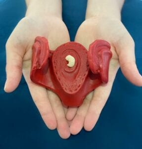
Most pregnancy apps will make you aware of the size comparison of your baby against a fruit/ vegetable/ animal or even baked goods, but rarely do you see images showing just how your baby would fit into the palm of your hand!
Take a look at this image which shows you the real size of your baby at 8 weeks!
At 8 weeks Dr Ushakov will be able to check for the heartbeat, date the pregnancy, check for single/multiple pregnancies. He will also check the structures of the pregnancy at this stage including the gestational sac, yolk sac and placenta.
During the Viability scan you will find out the estimated due date (Which will give you plenty of time to pack up that hospital bag and plan out your babymoon)!
If you are interested in having the viability scan at City Ultrasound, you can book our scans or give us a call on 02036872939 for more information
City Ultrasound is a pregnancy care centre based in the City of London in close proximity to Spitalfields Market. We specialise in the early diagnosis of congenital anomalies, and have more than 30 years of expertise in fetal medicine. Our clinic is equiped with the latest ultrasound equipment with the latest 3D/4D technology to produce outstanding quality of imaging for diagnostic purposes.
Our clinic has been recently refurbished to provide a relaxing environment for our patients who come in for early pregnancy reassurance. Watch this short Video Tour introducing our clinic and our staff!
Some media coverage of UCLH Spina Bifida unit on the BBC. Dr Fred Ushakov is a proud member of this clinic screening dozens of Spina Bifida cases a year from all over the UK. At City Ultrasound we screen for Spina Bifida from 10 weeks of pregnancy in our first trimester anomaly scan, also known as the Early Fetal Scan.
What is Spina Bifida?
Spina Bifida is a neural tube defect affecting the development of the spine and spinal cord of the baby. The neural tube is an embryonic structure that ends up forming the baby’s brain and spinal cord. There are three types of Spina Bifida; occulta, meningocele and myelomeningocele. Occulta (Latin for ‘hidden’) is the mildest form of Spina Bifida with symptoms including a hairy patch on the back or a dark spot on the back.
Myelomeningocele is the most severe anomaly and is commonly referred to as ‘open Spina Bifida’. The spinal canal remains open along several vertebrae in the back and exposing the baby’s tissues and nerves and forming a sac. Open Spina Bifida is associated with many other medical issues and is potentially life threatening for the baby. Some of the common complications associated with this type are: walking problems, orthopaedic issues, bowel issues, latex allergy, dermatological problems and accumulation of fluid in the brain.
According to the FMF, at 12 weeks the prevalence of Open Spina Bifida is 1 in 1,000 fetuses. In reality we believe that the condition is less prevalent in the UK but is still one of the most common neural tube defects. At City Ultrasound we screen for Spina Bifida from 10 weeks in our Early Fetal Scan.
Some of the risk factors for Spina Bifida include; folate deficiency (read our blog post on the Folic Acid), genetic history if neural tube defects, diabetes, certain medications and obesity.
One of Dr Fred Ushakov’s passions is teaching and training other professionals in the field of Fetal Medicine and the applications of ultrasound screening. Prior to the pandemic Fred lectured in countries such as the US, Australia, Italy, Sweden, Peru, Egypt, Romania, Ukraine and China, where the picture below was taken. Fred has also collaborated with GE Healthcare in live masterclass demonstrations in their Brussels HQ.
During the pandemic, Dr Ushakov was one of the pioneers to start broadcasting his lectures online. His lectures are mainly focused around his field of interest of early anomaly detection and the Early Fetal Scan. In the past year alone, over a thousand ultrasound professionals have attended his courses online.
Incredible shot of the Cerebellum at just 11 weeks in our recent instagram post!
This fetus is just 4cm long, but our advanced transvaginal probes are able to get this high resolution image of the cerebellum. Latin for ‘little brain’, the cerebellum is an important structure at the back of the human skull. The cerebellum is responsible for coordinating our voluntary movements, as well as motor skills, balance, coordination and posture.
It consists of two major structures namely the Cerebellar cortex and Cerebellar nuclei. Despite only being 10% of the human brain, the ‘little brain’ contains up to 80% of the brain’s neurons, mainly in the cortex. Neurons are important cells in the human body that are the building blocks of the nervous system.
You can see why it is important to check the development of the brain. Dr Fred Ushakov is a specialist in neurosonography and conducts a comprehensive examination of the brain with every scan from 11 weeks until the 3rd trimester.
The Umbilical Cord is an important structure, it is essentially a tube that connects the baby and the placenta. The tube usually starts developing from 5 weeks of gestation and grows until the beginning of the third trimester at ~28 weeks.
The umbilical cord consists of 3 blood lines; 2 arteries and 1 vein and basically acts as a supply line between the placenta and the baby. You can see why it is an extremely important structure as it delivers nutrients and oxygen to the baby and removes the baby’s “waste” materials.
The cord can grow up to 80cm, but the average is ~55cm, a longer cord can be associated with entanglement and possibly some complications during labour. Some potential complications in the pregnancy are related to the umbilical cord, such as Vasa Previa. The umbilical cord is cut at birth and the residual usually dries and falls off within 2-3 weeks.
In the video attached you can clearly see the umbilical cord attached from the placenta to the baby at just 12 weeks of gestation!
At City Ultrasound, the umbilical cord is just one of the important structures we check at any scan from 12 weeks. On our website, we have in-depth information about our process and types of scans, but if you do have any questions at all, please feel free to get in touch with us!
City Ultrasound is pleased to announce a long awaited relocation to Spital Square, just a few minutes’ walk from the iconic Spitalfields Market and adjacent to the famous Galvin La Chapelle restaurant. We are proud to be able to offer our patients a fantastic new clinic equipped with the latest ultrasound technology, a spacious and light waiting room and much more (sneak peek photos attached!)
The relocation is effective immediately, and we have closed our Finsbury Circus clinic permanently. The new location is conveniently located within 3 minutes’ walk from Liverpool Street Station. Additionally, paid street parking is available in front of our new building.
I would like to take this opportunity to thank you, our valued patients, for supporting our newly established service. When opening our doors for our first patient in April 2020, it took us almost a year to prepare for the launch. It has been a long and challenging journey but we would not have been able to expand and get to where we are now without your support.
Finally, if you would like to share your experience with City Ultrasound, I would really appreciate if you could kindly leave a comment on Google Reviews. We hope to see you at our new clinic soon!
In today’s video you can clearly see the baby resting in the Amniotic Sac at about 11 weeks and even see its brain!
The amniotic sac is a pair of membranes (basically a filter that lets some things pass but stops others) where the baby is developing. The inner membrane is known as amnion, while the outer membrane is known as chorion. The amnion is filled with a fluid suitably called amniotic fluid which transfers essential substances such as oxygen from the umbilical cord to the baby. The chorion contains the amnion and is part of the placenta.
During labour, the amniotic sac breaks resulting in the ‘water breaking’ when the baby is ready to come out. In some cases, the amniotic sac doesn’t break during labour and the baby is born in a transparent bubble, this is quite rare and only happens in 1 in 80,000 cases- known as en caul birth!
At City Ultrasound, the amniotic sac is just one of the important structures we check at any scan from 8 weeks. On our website, we have in-depth information about our process and types of scans, but if you do have any questions at all, please feel free to get in touch with us!
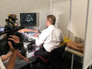
What is a transvaginal scan (TVS)? “Transvaginal” means an internal examination through the vagina. TVS probes have extraordinary resolution and may provide unique information regarding the fetal anatomy, placenta, womb, cervix and other important structures.
Technically, examination by TVS during pregnancy is identical to gynaecological ultrasound. The special high-resolution probe is aseptically cleaned, covered with a sterile cover (like a condom) and sterile lubricating gel. The transducer inserted is no larger than a finger. It is gently passed into the vagina to generate images of the baby, while you lay on your back. This may cause some slight discomfort but should not cause any pain. It is important you let us know if you have a latex allergy, in which case we will use latex-free probe covers.
The operator performing the transvaginal scan in pregnancy must have a high level of expertise and experience. TVS scan represents a significant challenge for the doctor or sonographer because of difficulties in obtaining correct images. As such, there are only a few specialists in London performing transvaginal assessment of the early fetus, including Dr Ushakov.
Please empty your bladder immediately before the transvaginal scan. Even a small amount of urine in the bladder will change the position of the womb and will reduce the quality of image. A trained chaperone is also available and present for internal examinations and may be requested for any of our other scans.
Is TVS safe? TVS uses the same safe ultrasound waves as transabdominal scans to obtain the images of the baby and womb. The probe cannot get into direct contact with the fetus. Normally, due to better resolution the time of TVS imaging is shorter in comparison to a transabdominal scan (approximately 10 minutes). We use rigorous aseptic technique for preparation of the probe and there is no risk of infection.
Visit our Instagram account for comparison of transvaginal (TVS) & transabdominal scans for the SAME pregnancy. Can you see how much better the resolution of the TVS scan (1st video)? This is generally the case for ultrasound scans up to 11weeks when the fetus is still very small, less than 1.6 inch!