
Tag: fertility

Hertility x LPC partnership

Minimalist flat design illustration of fertility and pregnancy ultrasound partnership between specialists London Pregnancy Clinic and Hertility.

Hertility x LPC partnership:

Bringing Fertility and pregnancy ultrasound support for women

  • Published

  • Last Modified

Tags

Discover the transformative partnership between Hertility and London Pregnancy Clinic, revolutionising the way you access fertility and pregnancy ultrasound services. From early pregnancy to specialised scans, our collaboration ensures you receive world-class care without the wait, at a price you can afford.

In an exciting development for expectant mothers and those on their fertility journey, Hertility has partnered with us, London Pregnancy Clinic (LPC), transforming access to reproductive healthcare. Understanding the emotional toll of waiting for your first pregnancy scan, we’ve come together to offer you world-class scanning facilities, ensuring early reassurance through ultrasound without the long wait. It promises to bridge the gap between fertility understanding and pregnancy care, offering an integrated approach to women’s health that is both convenient and comprehensive. Both fertility and pregnancy ultrasound are very important and we are going to unpack what we can offer.

Who is Hertility?

In 2020, Hertility was conceived with a distinct vision by Dr. Helen O’Neill, Dr. Natalie Getreu, and Deirdre O’Neill. It sprang from a realisation that women’s health complexities were often overlooked, with conditions remaining enigmatic within the healthcare system. Hertility emerged as a beacon, championing the cause of bringing reproductive science into the hands of women globally.

Tired of the gaps in women’s healthcare, Hertility was born out of necessity. It became clear that the data underpinning women’s health was not only insufficient but also largely based on male physiology, leading to a paradigm shift in approach. Dr. O’Neill, through her fifteen years in academia, recognised the urgency for tailored datasets that reflect the true nature of female health concerns. Hertility is not just a company; it’s a movement towards empowering women with the knowledge to make informed decisions about their health and life choices.

Why was Hertility Created?

The inception of Hertility was motivated by the profound desire to address the elephant in the womb – the inherent complexities of female reproductive health. With a staggering 60% of women grappling with hormone and gynae issues and the diagnosis of fertility disorders stretching up to eight years, Hertility’s founders saw the dire need for change. By empowering women with accessible, research-led health assessments, Hertility is breaking barriers and shining a light on the path to understanding and managing women’s health effectively.

Hertility’s resolve to be research-led has set a new standard in women’s healthcare. It stands firmly on the foundation of clinical trials and extensive data collection, ensuring every insight provided is robust and actionable. Hertility’s comprehensive at-home hormone and fertility tests are more than just diagnostics; they are a clarion call to acknowledge and act on women’s reproductive health needs. The company’s commitment to sharing their findings reflects their ethos of transparency and education, making Hertility a true vanguard in the realm of women’s health.

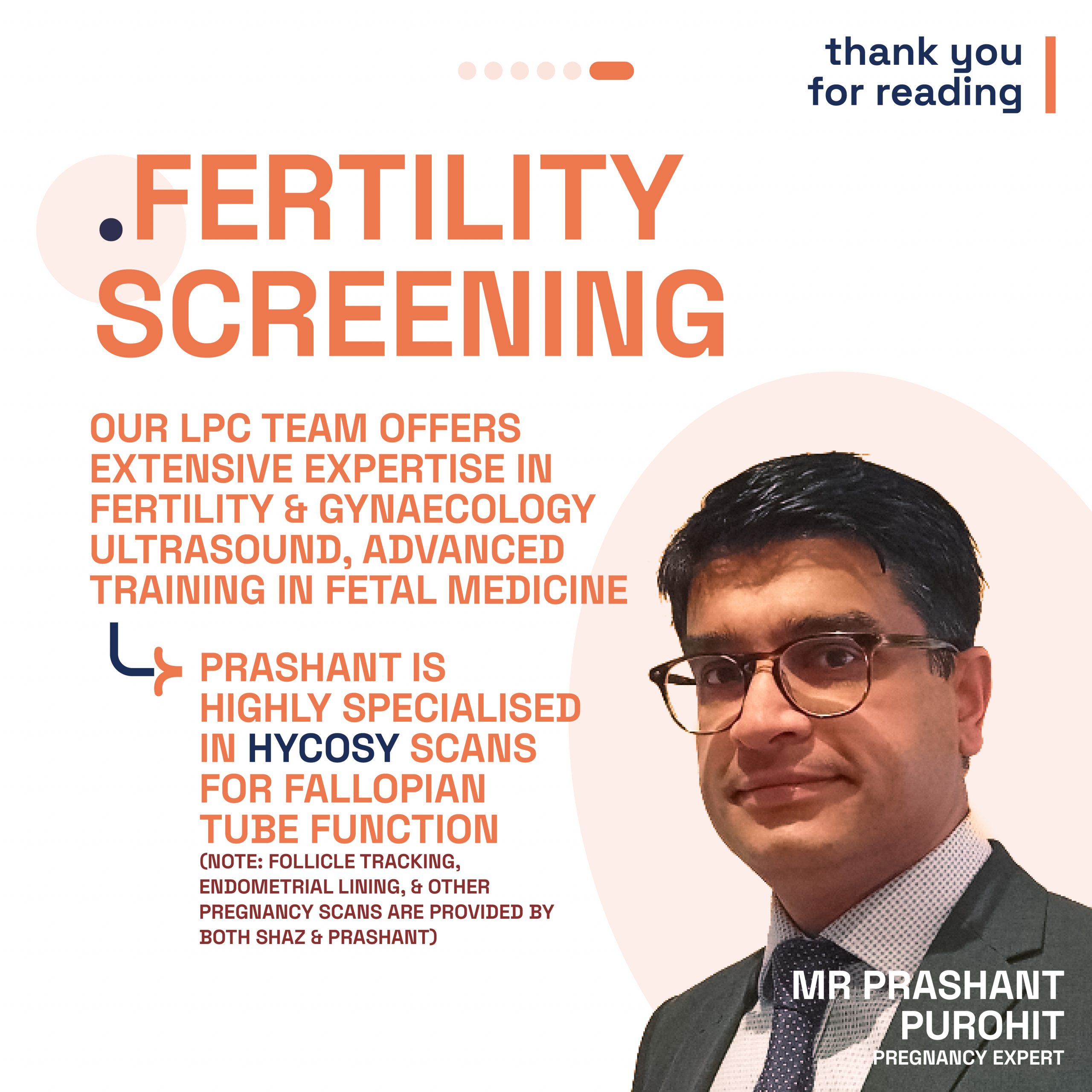
Why Hertility and LPC?

Hertility is at the forefront of women’s health, offering at-home hormone and fertility test kits designed to empower women with knowledge about their reproductive health. Their comprehensive approach evaluates various factors, including hormonal balance, ovarian reserve, and potential reproductive health conditions, to provide actionable insights.

Understanding the importance of seamless care, Hertility’s partnership with LPC is a natural progression. LPC’s reputation for excellence in fertility and pregnancy ultrasound complements Hertility’s mission, providing a continuum of care that supports women from fertility assessment through to pregnancy. From fertility screening to anomaly scan we provide a large variety of services for women from our London clinic.

  • Fertility and pregnancy ultrasound services at  London Pregnancy Clinic - Viability scan

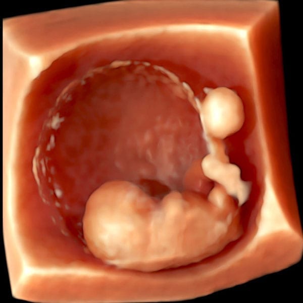
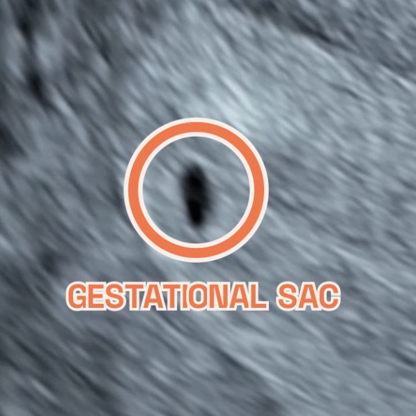
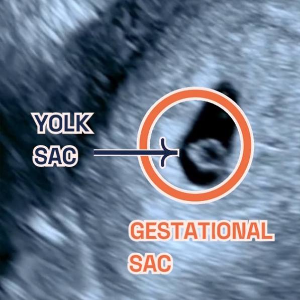
    Viability scan 3D image

  • Image showing what would an ultrasound show at 6 weeks of pregnancy. You can see the fetal pole, gestational sac and yolk sac. It shows an Ultrasound image by London Pregnancy Clinic that shows the gestational sac and how small it is - for educational purpose.

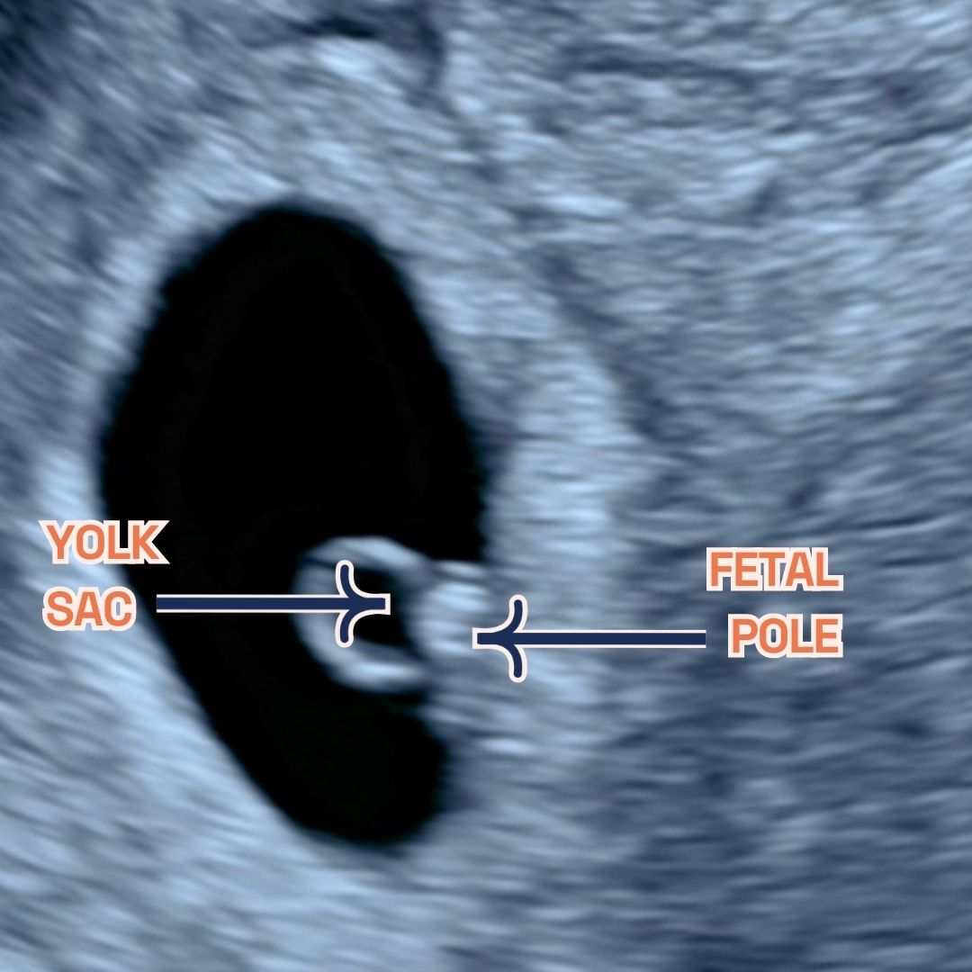
    You can see the fetal pole, gestational sac and yolk sac at week 6.

Fertility and pregnancy ultrasound

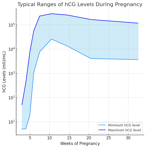
  • Early Pregnancy Scans: Available from 6 weeks, these scans offer early reassurance, confirming the presence of a gestational sac and fetal heartbeat. The first scan we offer is called the viability scan (or the 6-week scan).
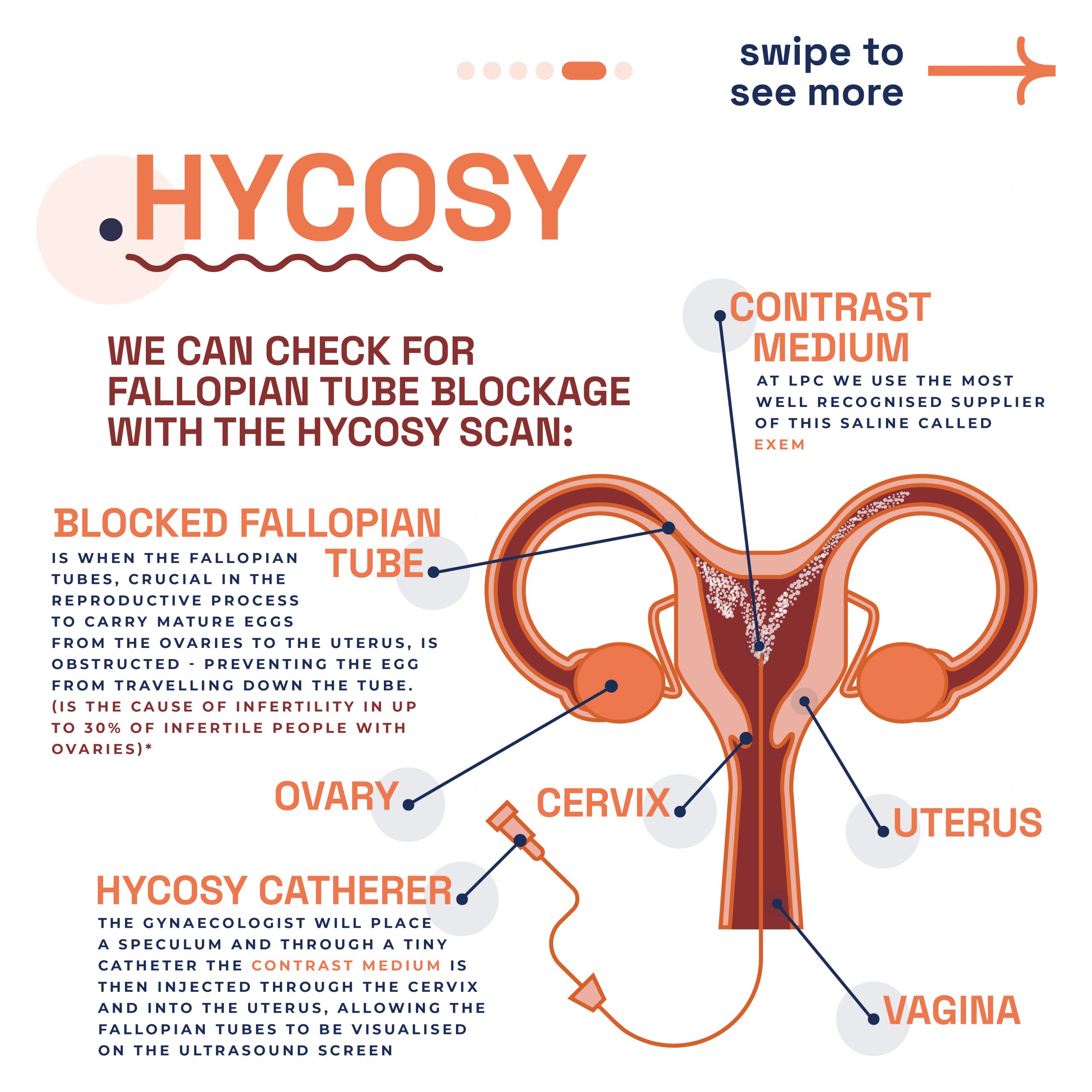
  • HyCoSy Scans: For those investigating fertility concerns, HyCoSy scans assess tubal patency, crucial for understanding potential fertility issues.
  • General, Anomaly, and Early Fetal Scans: A comprehensive range of scans ensures that every aspect of your pregnancy is monitored, from general health checks to detailed anomaly scans. We specialise in NIPT and 10 Week Scan, which is our signature early pregnancy scan.

All scans at LPC are conducted by consultant-level doctors or specialist sonographers in fetal medicine, gynaecology, and fertility. Overseen by Dr Fred Ushakov, a renowned specialist in fetal medicine and obstetrical ultrasound, our team guarantees the highest standard of care.

Why Wait? Early Scans Available from 6 Weeks

The first few weeks of pregnancy are a profound journey filled with anticipation and, sometimes, anxiety. Traditionally, expecting mothers wait until the 11 to 14-week mark for their first scan, a period filled with unanswered questions and needless worries. Recognising the importance of early reassurance, LPC opens its doors to Hertility customers for pregnancy scans starting as early as 6 weeks. Our goal? To provide you with the peace of mind that comes from knowing your pregnancy is progressing as expected, as soon as possible.

If you’re navigating the journey of fertility and pregnancy, let Hertility and LPC guide you to a path of informed, supported, and comprehensive care. You can check out our fertility and pregnancy ultrasound services!

Final thoughts

Hertility’s partnership with the London Pregnancy Clinic marks a defining moment in women’s health. With Hertility’s research-driven methodology and our fertility and pregnancy ultrasound services, we hope to provide a holistic approach for all women. Our combined effort provides an answer to the calls for early and accessible pregnancy scans, ensuring that expectant mothers receive the support and assurance they need, precisely when they need it. This collaboration goes beyond convenience; it is a powerful statement that women’s health deserves prioritisation and respect.

Latest Stories

Continue reading

What is a Fallopian Tube?

Illustration detailing the expert fertility assessment of fallopian tubes offered at London Pregnancy Clinic.

Fallopian Tubes: all you need to know

Discovering the importance of fallopian tubes for fertility.

  • Published

  • Last Modified

Tags

Explore the critical role of fallopian tubes, or uterine tubes, in pregnancy and fertility. This post delves into their anatomy, function, and common issues affecting fertility, providing essential insights for expectant mothers and those planning pregnancy.

Welcome to our comprehensive guide on the fallopian tubes, an essential component of the female reproductive system. We also explore common pathologies that affect these tubes, leading to infertility issues and complications like ectopic pregnancies. Our esteemed in-house gynae team, brings their extensive knowledge and expertise to this discussion, providing invaluable insights into the significance of maintaining fallopian tube health. Join us as we unfold the mysteries of the fallopian tubes and their vital function in the miracle of life.

What are Fallopian Tubes

In the female body, the pelvis houses two vital structures known as fallopian tubes. One on either side of the uterus. These tubes act as pathways, connecting the ovaries to the uterus. Each fallopian tube is made up of four distinct parts: the infundibulum, fimbriae, ampulla, and isthmus. The fimbriae are delicate, finger-like extensions that gently guide the egg from the ovary into the tube. 

The Role of Fallopian tubes in Pregnancy

Moreover, fallopian tubes are critical in the earliest stages of pregnancy, integral to the reproductive process, and serve a trio of critical functions. They are the conduits for egg transportation from the ovaries to the uterus and the venue where fertilisation typically occurs. Post-fertilisation, these tubes facilitate the journey of the fertilised egg to the uterus, setting the stage for implantation and the progression of pregnancy. The health and integrity of the fallopian tubes are paramount for successful conception as they are closely linked to fertility. Problems with these tubes are a common reason why some women may have difficulty getting pregnant.

Uterine Tube Pathologies & Infertility

According to a report, nearly one-third of infertility cases are attributed to issues with the fallopian tubes, including various pathologies like inflammation and obstructions. Damage to the tube’s cilia can hinder the movement of sperm or egg, contributing to infertility. Additionally, sexually transmitted infections are a known cause of these tubal issues. Let’s dive into the specific pathologies that can manifest.

Salpingitis

Salpingitis, an inflammation in the fallopian tubes, often forms part of wider pelvic inflammatory diseases (PIDs). Its hallmark is a thickened tube in the isthmus area, known as salpingitis isthmica nodosa. This condition can resemble endometriosis, potentially leading to fertility blockages or raising ectopic pregnancy risks.

During diagnosis, doctors first look for tenderness and swelling. They may conduct blood and urine tests to spot infection indicators. Swab tests from the vagina and cervix help identify specific bacterial infections. Ultrasound scans of the fallopian tubes and reproductive tract are common. A crucial test, the hysterosalpingogram, uses a special X-ray and dye through the cervix to detect tubal blockages.

Untreated, salpingitis can cause long-term fertility issues and increase ectopic pregnancy chances. Symptoms vary, including lower abdominal pain, nausea, and fever; sometimes, no symptoms appear initially. Antibiotics typically treat this condition, but severe cases might require surgery or IVF. Seeking medical advice early is vital to prevent serious outcomes. Early detection and treatment significantly aid in managing salpingitis effectively.

Fallopian Tube Blockage & Narrowing

Addressing fallopian tube obstructions can significantly boost pregnancy chances. These blockages may develop in various parts of the tube – proximal, distal, or mid-segmental. Testing the tubes’ full functionality presents challenges, yet assessing their patency (openness) is possible with hysterosalpingography, laparoscopy with dye, or hysterosalpingo contrast sonography (HyCoSy). During surgical evaluations, specialists examine the tubes and introduce a dye, such as methylene blue, into the uterus to test its passage through the tubes when the cervix is closed.

Research published in PubMed indicates a notable prevalence of tubal obstruction: 19.1% in primary infertility cases and 28.7% in secondary infertility scenarios. The study highlights cornual blockage’s predominance in primary infertility, whereas complete hydrosalpinx and peritubal adhesions appear less frequently. Due to the frequent link between tubal disease and Chlamydia infection, Chlamydia antibody screening is now a cost-effective approach for identifying possible tubal pathologies.

At London Pregnancy Clinic, we conduct the HyCoSy procedure. This test evaluates fallopian tube functionality by injecting a special dye and monitoring its flow. It also assesses the womb for abnormalities affecting pregnancy implantation. Additionally, we use dye and ultrasound scans to check for fallopian tube blockages or abnormalities. Discover more through the slider below, offering further insights into our advanced diagnostic techniques.

  • Graphic explaining the expert fertility assessment of fallopian tubes offered at London Pregnancy Clinic.

    Fallopian Tubes

  • Educational diagram of female reproductive anatomy focusing on the fallopian tubes provided by London Pregnancy Clinic.

    Fallopian tube diagram

  • Illustration detailing the phases of the menstrual cycle as part of fertility education by London Pregnancy Clinic.

    Menstrual Cycle

  • Infographic showing statistics on blocked fallopian tube prevalence in women, as part of London Pregnancy Clinic's fertility resources.

    Stats on blocked fallopian tube

  • Explanation of the HYCOSY scan procedure for checking fallopian tube blockage, offered by London Pregnancy Clinic.

    HyCoSy

  • Image of Mr. Prashant Purohit with text highlighting London Pregnancy Clinic's expertise in fertility and gynaecology ultrasound.

    Mr. Prashant Purohit specialist gynaecologist

Ectopic Pregnancy

Statistics and Locations of Ectopic Pregnancies: Ectopic pregnancies, which occur in about 1%-2% of all pregnancies, predominantly implant in the fallopian tubes, with over 98% of cases occurring there. Within the fallopian tubes, the distribution of ectopic pregnancies varies: approximately 75% happen in the ampullary segment, 13% in the isthmic segment, and around 12% in the fimbrial segment. These statistics highlight the critical nature of monitoring tubal health.

Fertilisation and its Relation to Ectopic Pregnancies: It’s important to note that fertilisation typically occurs in the ampulla, the same section of the fallopian tube where the majority of ectopic pregnancies are found. This correlation emphasises the need for awareness and early detection in cases of suspected ectopic pregnancy, as the ampulla is a common site for both normal fertilisation and ectopic implantation.

Understanding the statistics and locations of ectopic pregnancies in the fallopian tubes is crucial. It not only sheds light on the commonality and risks associated with these pregnancies but also underscores the importance of early ultrasound pregnancy scans for detection and intervention. The earlier you scan the quicker we can detect an ectopic pregnancy meaning that we can begin this from week 6 of pregnancy.

Pregnant with Just One Fallopian Tube?

Typically, women possess two fallopian tubes for transporting the mature egg from the ovary to the uterus, where it may encounter sperm. However, a single healthy tube can efficiently undertake this task. Remarkably, up to 85% of women aged 22-28 with only one tube successfully conceive within two years of trying, even following an ectopic pregnancy.

If you have a blocked tube due to scarring, trauma, or a previous ectopic pregnancy, your pregnancy prospects remain normal, considering other health factors. Loss of a fallopian tube might result from pelvic surgery due to infection, tumours, or a past ectopic pregnancy. In some instances, women are naturally born with just one tube. Still, pregnancy is achievable with one tube, provided you have a functioning ovary, you’re ovulating and the fallopian tube is in good health.

If you’re struggling to conceive or have a history of fallopian tube issues, it’s wise to consult your gynaecologist or a reproductive endocrinologist. They can offer a thorough evaluation to guide your next steps. Remember, one healthy fallopian tube can effectively support your journey to motherhood.

Conclusion

Mr. Prashant Purohit, our dedicated in-house gynaecologist at London Pregnancy Clinic, emphasises the need for awareness and timely medical consultation to address these concerns. His expertise in managing these conditions reflects our clinic’s commitment to providing comprehensive care and support to women facing fertility challenges. Understanding the complexities of the fallopian tubes is not just about medical knowledge; it’s about empowering women with the information they need to make informed decisions about their reproductive health. We encourage you, readers to stay vigilant about their gynaecological well-being and seek expert guidance when needed. Remember, early detection and treatment are key to overcoming obstacles on the path to motherhood.

Latest Stories

Continue reading

Week 5 of Pregnancy

Graphic of Week 5 of pregnancy. An illustration of a baby in the ultrasound machine.

Week 5 of your Pregnancy

Discovering your baby’s progress since week 4

  • Published

  • Last Modified

Tags

Having journeyed through the early whispers of Week 4, we now step into the heart of your pregnancy story: Week 5. It’s a chapter filled with vibrant growth and transformation. Imagine your little one, still as tiny as an apple seed, beginning a significant journey into the embryonic period.

Week 5 of pregnancy, it’s all about growth and transformation. Your little one is busy developing, cell by cell, quickly growing from the size of a poppy seed in week 4 (watch video here) to an apple seed. We’ll explore these changes together, learn how to take care of yourself during this time and get a peek into your baby’s development with early ultrasounds. So, let’s dive into Week 5, where every day brings something new and amazing in your pregnancy journey. So let’s navigate the changes unfolding, how to care for yourself, and what to expect from medical check-ups.

Key Concepts: embryonic period

During Week 5 of your pregnancy, a crucial phase begins as your baby, the size of an apple seed, enters the embryonic period. This week marks the foundation for major organs and systems. Hormonal changes, especially increases in progesterone and human chorionic gonadotropin (hCG), are vital in supporting this stage. These hormones maintain the uterine lining and ensure the continuation of progesterone production, crucial for pregnancy health. As a result, you might notice early symptoms like fatigue and nausea. Understanding these changes is key to navigating the early stages of your pregnancy journey.

Pregnancy Checklist at 5 Weeks

While it’s still early in your pregnancy, there are some important steps you can take:

  • Confirm your pregnancy with a test if you haven’t already.
  • Schedule your first prenatal appointment to discuss your health and pregnancy. At London Pregnancy Clinic it’s called the Viability/Dating Scan. Use our pregnancy calculator to work out the best time for your scan.
  • Begin focusing on a pregnancy-safe diet and lifestyle, emphasising foods rich in folic acid and essential nutrients.

Developmental Milestones: Week 5

Growth of the Gestational Sac: This week, the gestational sac, housing your developing baby, continues to grow. It’s a clear sign of your pregnancy’s progression, providing a protective and nourishing environment for the embryo.

Yolk Sac Development: The yolk sac plays a crucial role at this stage, providing essential nutrients before the placenta is fully operational. It also contributes to the early stages of the circulatory system by producing blood cells, supporting the developing embryo’s needs. This process is essential for supporting the growing needs of your embryo and is closely tied to the growth of the gestational sac.

Heart Development: Now your baby’s heart begins its incredible journey. Initially emerging as a simple tube-like structure, it’s preparing to pulsate and evolve into a sophisticated four-chambered heart in the coming weeks. Although it’s not beating just yet, this early stage sets the stage for one of life’s most vital rhythms.

Brain and Spinal Cord Formation: Alongside the heart, the neural tube, which will become the brain and spinal cord, starts to take shape. This tube forms the bedrock of the entire nervous system, integral for everything from physical movements to cognitive functions. It’s a complex process that lays the foundation for your baby’s future mental and physical capabilities.

Emergence of Other Organs: Week 5 also marks the beginning of other organ formations. Organs like the kidneys and liver start to form, albeit in their very preliminary stages much like the heart.

What is seen on the Ultrasound: Week 5?

During the Week 5 ultrasound, expect to see the gestational sac (which has dramatically increased in size since Week 4), the first structure visible on an ultrasound, indicating your pregnancy is progressing well. This small, fluid-filled sac is where your baby will develop. Inside it, the yolk sac may also be visible, which is crucial for providing nutrients to the developing embryo and for blood cell production in these early stages. However, it’s important to remember that at this stage, the embryo itself and its heartbeat are usually not yet detectable.

It’s common not to see the embryo or hear a heartbeat during a Week 5 ultrasound. The embryo is still very small, and the heartbeat typically becomes detectable around the 6th or 7th week of pregnancy. While we know it’s tempting to have an early ultrasound, we at London Pregnancy Clinic recommend waiting until at least week 6 for your first scan. This crucial ultrasound provides a tangible glimpse into your pregnancy, offering reassurance and joy as you embark on this extraordinary journey.

  • Gestational sac at 4 weeks of pregnancy as seen on ultrasound scan.

    Gestational sac at 4 weeks of pregnancy as seen on ultrasound scan.

  • Image showing what would an ultrasound show at 5 weeks of pregnancy. It shows an Ultrasound image by London Pregnancy Clinic that shows the gestational sac and the newly developed yolk sac - for educational purpose.

    Gestational sac and yolk sac at 5 weeks of pregnancy as seen on ultrasound scan.

Thinking ahead: Week 6?

Looking forward to Week 6, expect continued development of your baby’s facial features. You may also start experiencing more pronounced pregnancy symptoms like nausea and fatigue. It’s a time to start preparing both mentally and physically for these changes.

Week 5 of Pregnancy FAQs


  • How common is it for my baby to have a heartbeat at 5 weeks?

    Your baby is an embryo (not a fetus just yet) and can have a heartbeat at 5 weeks of pregnancy, but it is not very common. The fetal heartbeat usually starts to beat between 5 and 6 weeks gestation, and it may be detectable via ultrasound around this time.

    Here are some reasons why the heartbeat might not be detected on an ultrasound at 5 weeks:

    • The embryo is too small.
    • The heartbeat is too faint.
  • What if there is no yolk sac at 5 weeks?

    Patients have asked us why is there no sac seen when 5 weeks pregnant. The yolk sac is usually visible on an ultrasound between 5 and 6 weeks gestation. If there is no yolk sac visible on an ultrasound at 5 weeks, it could be a sign of a miscarriage. 

    However, it could be a sign that the estimated gestational age is wrong. This could happen if your last menstrual period was inaccurate or if you were ovulating later than you thought.

    In this case, we may recommend a follow-up ultrasound in a week or two to see if the yolk sac has developed.


Conclusion

Week 5 of your pregnancy is a time of rapid and incredible changes. While you might start feeling different physically and emotionally, remember, these are signs of your baby’s healthy development. Stay informed, take care of yourself, and prepare for the exciting journey ahead. If you have any concerns, our team at London Pregnancy Clinic is always here to guide you.

Stay tuned on what your baby’s week-by-week growth will look like on our blog.


Latest Stories

Continue reading

Week 4 of Pregnancy

Graphic of Week 4 of pregnancy. An illustration of a zygote in the uterus.

Week 4 of your Pregnancy

Your Baby’s Remarkable Beginning

  • Published

  • Last Modified

Tags

This blog post is part of a series that breaks down early pregnancy week by week. Today we will be talking about Week 4 of your pregnancy journey! Within this article, we’ll delve into the astonishing advancements occurring during this initial phase of pregnancy. Although your baby remains incredibly minuscule, measuring at no more than 1 millimeter (similar to a poppy seed), a realm of growth and transformation is already well underway.

Key Concepts: Gestational Weeks vs. Post-Conception Weeks

Understanding the difference between gestational weeks and post-conception weeks is crucial for accurately tracking the progress of your pregnancy.

Gestational Weeks: Gestational age refers to the age of the pregnancy and is measured from the first day of the mother’s last menstrual period (LMP). This is the standard method used by healthcare providers to track pregnancy. For example, at 4 weeks gestation, the embryo is about 2 weeks post-conception. Gestational age is crucial for determining the due date and monitoring the baby’s development.

Post-Conception Weeks: Post-conception age, also known as embryonic age, is measured from the time of conception. It provides a more precise timeline of the baby’s development. For instance, at 4 weeks post-conception, the embryo is undergoing significant developments, such as the formation of the placenta and the beginning of the amniotic sac.

Pregnancy Checklist at 4 Weeks

While it’s still early in your pregnancy, there are some important steps you can take:

  • During the initial 4 weeks of pregnancy, you may not experience noticeable symptoms. To verify your pregnancy, consider taking a pregnancy test.
  • Once you’ve confirmed your pregnancy you may think about your first prenatal visit with your doctor, even though it may not occur for another couple of weeks. With us, this would be our Viability/Dating Scan. During this visit, our specialists, like Miss Shaz Khojasteh, will check for single/multiple pregnancies, date the pregnancy and check the main structures of the gestational sac. We believe early pregnancy scans are essential for peace of mind and early reassurance for parents. 
  • Focus on adopting healthy diet and lifestyle habits. Say goodbye to smoking and alcohol, opt for nutritious foods, and stay well-hydrated.
  • The NHS provides valuable recommendations for expectant mothers, including considering the importance of taking supplements during pregnancy. Additionally, it’s essential to recognise how emotional changes that often accompany pregnancy can impact your relationships.

Developmental Milestones: Week 4

By Week 4, your baby develops from being a single-cell zygote to a blastocyst. Rapid cell division shapes the future. At this stage, your embryo completes its journey from the fallopian tube to the uterus, burrowing into the uterine lining. Half becomes your future child, and the other forms the placenta—a vital nutrient carrier.

The amniotic sac, often called the “bag of waters,” forms around the embryo, along with the yolk sac, important for your baby’s digestive system.

Your embryo now consists of three unique cell layers:

  • The endoderm—nurturing your baby’s digestive system, liver, and lungs.
  • The mesoderm—laying the foundation for your baby’s heart, sex organs, bones, kidneys, and muscles.
  • The ectoderm—shaping your baby’s nervous system, hair, eyes, and outer skin layer.

What is seen on the Ultrasound: Week 4?

One of the common queries we receive at the London Pregnancy Clinic is whether having an ultrasound at 4 weeks gestation is a requirement. At this stage, around the fourth week of pregnancy, significant developments are occurring. The blastocyst is in the process of dividing into an embryo and placenta. However, it’s crucial to understand that an ultrasound of your uterus during this early stage will typically reveal what appears to be a minuscule dot known as the gestational sac, and it’s important to note that a heartbeat is not typically detectable at this early point of development. Detecting a more advanced pregnancy may require waiting until a later stage.

Thinking ahead: Week 5?

The next steps in your pregnancy journey involve preparing for your first ultrasound appointment. It’s completely normal to eagerly anticipate your first ultrasound, but it’s essential to be aware that at 4 and 5 weeks of gestation, the gestational sac is typically too small to be easily visible on the scan. For a clearer image of your pregnancy, it is generally advisable to wait until around 6 weeks or later, when the development progresses. However, it’s worth noting that in certain cases, skilled sonographers with extensive experience may be able to discern subtle indications of pregnancy by closely observing changes in the uterine lining. You can use our Pregnancy Calculator to work out your optimal scan date range by entering your Last Menstrual Period(LMP).

Week 4 of Pregnancy FAQS?


  • Is 4 weeks pregnant too early to test

    At 4 weeks, it’s not too early to take a pregnancy test. Most home pregnancy tests are designed to be sensitive enough to detect the pregnancy hormone, human chorionic gonadotropin (hCG), in your urine by this time. This is typically around the time your period is due, which is when hCG levels are sufficiently high to indicate pregnancy.

    While you can confirm pregnancy with a test and even start calculating your due date, it’s indeed too early for an ultrasound scan. At 4 weeks, the baby is very small, and significant developmental milestones like the heartbeat, which typically becomes detectable around 6 weeks of gestation, have not yet occurred. Therefore, while a home pregnancy test can provide early confirmation, an ultrasound scan at this stage wouldn’t offer much information. For more detailed and visual insights into your baby’s development, waiting until at least the 6th week or later is advisable when the heartbeat and other embryonic structures start to become visible.

  • What to do when you’re 4 weeks pregnant?

    • Confirm Your Pregnancy: A home pregnancy test can confirm your suspicion of being pregnant. It’s best to take the test after you’ve missed your period for the most accurate result.

    • Schedule a Doctor’s Appointment: Even though it’s early, getting in touch with a healthcare provider is a good idea. They can confirm your pregnancy through a blood test and start guiding you through prenatal care.

    • Document Your Journey: Consider starting a pregnancy journal or diary to record your experiences, thoughts, and feelings. This can be a wonderful keepsake for the future.

    • Rest and Relax: Early pregnancy can bring fatigue and other symptoms. Ensure you’re getting enough rest and practising stress-relief techniques.

  • What does 4 weeks pregnancy look like?

    At 4 weeks of pregnancy, external changes to your body are typically minimal and not yet noticeable to others. However, internally, a remarkable series of events is unfolding. Your embryo, though only about the size of a poppy seed, is rapidly developing. This tiny cluster of cells is already beginning the incredible process of forming what will become vital organs and structures.


Conclusion

Week 4 of your pregnancy signifies the remarkable commencement of your baby’s journey. While ultrasound visuals may not reveal much at this early stage, rest assured that a realm of development is quietly unfolding within your body. Join us as we look forward to exploring the upcoming stages of your pregnancy journey, complete with the changes and milestones that await.

It’s crucial to remember that every pregnancy is unique. Therefore, we recommend consulting your healthcare provider for tailored guidance and attentive care during this special period. Our team is here to provide support throughout your exciting pregnancy journey!

Once again, heartfelt congratulations on your pregnancy. Stay tuned for further updates as we continue to monitor your baby’s week-by-week growth.


Latest Stories

Continue reading

A Comprehensive Guide to AMH Blood Test: Its Significance and Connection to IVF

Ever wondered how a simple blood test could offer a glimpse into your fertility? In this blog post, we’ll delve into one of the most crucial hormones affecting fertility—the Anti-Mullerian Hormone (AMH). If you’ve heard of this hormone and wondered what role it plays in fertility or IVF treatments, you’re in the right place.

What is the AMH Blood Test?

An AMH (Anti-Mullerian Hormone) blood test measures the levels of AMH in a woman’s blood. Produced by the cells of the ovarian follicles, AMH serves as a reliable marker of ovarian reserve, effectively indicating a woman’s fertility potential. This test aids clinicians and fertility specialists in predicting a woman’s response to fertility treatments, particularly in vitro fertilisation (IVF).

What is the Anti-Mullerian Hormone (AMH)?

The Anti-Mullerian Hormone, first discovered by Alfred Jost in the 1940s, plays a crucial role in the sexual development of foetuses. AMH was initially known for its role in male sexual development, but subsequent research has unearthed its pivotal part in female fertility. In women, the hormone is released by the antral follicles, the small sacs in the ovaries that hold the eggs.

Why is the AMH Blood Test Important?

As AMH levels correspond to the number of remaining eggs in the ovaries, the AMH blood test offers a reliable gauge of a woman’s ovarian reserve. However, it’s essential to understand that AMH levels do not directly correlate with the quality of eggs.

Low AMH levels might indicate a diminished ovarian reserve, which could be a concern for women attempting to conceive naturally or through IVF. In contrast, higher AMH levels may suggest conditions like Polycystic Ovary Syndrome (PCOS).

When is an AMH Blood Test Required?

The AMH test may be recommended in several circumstances:

  1. Evaluating Ovarian Reserve: AMH levels help assess the number of eggs remaining in the ovaries. The test may be suggested for women approaching their mid-30s, those with a family history of early menopause, or those who’ve previously undergone chemotherapy or pelvic surgery.
  2. Diagnosing PCOS: Elevated AMH levels may be indicative of PCOS, a common condition affecting a woman’s ovaries and ovulation.
  3. Determining IVF Success: Before starting IVF treatment, clinicians may order an AMH test to better predict how the ovaries will respond to the fertility drugs used to stimulate egg production.

Connection between AMH Levels and IVF

The AMH blood test holds significant value in the field of assisted reproductive technology, especially IVF. It helps clinicians devise personalised treatment strategies for their patients.

For women with low AMH levels, special considerations may be needed during the stimulation phase of IVF. These women often respond less to ovarian stimulation, leading to a smaller number of eggs retrieved, which can affect the success rate of IVF. Conversely, women with high AMH levels might be at risk for Ovarian Hyperstimulation Syndrome (OHSS), a condition that can occur when the ovaries are overly stimulated by fertility drugs.

When is the Best Time to Have an AMH Blood Test?

An AMH blood test can be performed at any time during a woman’s menstrual cycle, as AMH levels remain relatively constant throughout. However, many fertility clinics might suggest the test as a part of an initial fertility assessment or prior to beginning fertility treatments like IVF.

In Conclusion

The AMH blood test serves as a potent tool in assessing a woman’s ovarian reserve and planning personalised fertility treatments. Whether you’re attempting to conceive naturally, preparing for IVF, or just wish to know more about your fertility, an AMH test could offer invaluable insights. Always consult with a fertility specialist to discuss your options and understand what’s best for your individual circumstances.

Discover more about the Blood Tests we offer at London Pregnancy Clinic.

Pregnancy Testing – Deep Dive into the Beta HCG Test

Pregnancy tests aim to provide a simple yet effective method of determining if you’re pregnant. There are two primary types of pregnancy tests available: urine tests and blood tests. While urine tests are more commonly used due to their convenience and over-the-counter availability, blood tests, specifically the Beta HCG (Human Chorionic Gonadotropin) test, offer a more detailed insight into the pregnancy. Here’s an in-depth look at the Beta HCG pregnancy test, how it differs from urine tests, its accuracy, uses, and how to interpret the results.

What is a Beta HCG Pregnancy Test?

A Beta HCG test is a blood test used to detect pregnancy. Human Chorionic Gonadotropin (HCG) is a hormone produced by the placenta after implantation. The beta subunit of HCG can be detected in the blood serum of expectant mothers approximately 11 days after conception; HCG can be detected in the urine 12-14 days after conception.

There are two types of HCG tests:

  1. Qualitative HCG test: This test simply detects the presence of HCG in the blood. It gives a “yes” or “no” answer to the question, “Are you pregnant?” Doctors often order this test to confirm pregnancy.
  2. Quantitative HCG test (Beta HCG): This test measures the specific level of HCG in the blood. It can detect even very low levels of HCG, making it a highly accurate test.

How Does a Beta HCG Test Differ from Urine Tests?

While both urine pregnancy tests and Beta HCG tests aim to detect the presence of HCG, there are distinct differences:

  1. Method of Testing: Urine tests can be done at home, while Beta HCG tests require a blood sample taken in a healthcare setting.
  2. Sensitivity: Beta HCG tests are more sensitive than urine tests and can detect lower levels of HCG. This means they can confirm pregnancy earlier than urine tests — typically around 11 days after conception.
  3. Information Provided: While urine tests only provide a positive or negative result, a Beta HCG test can provide more information about the pregnancy, like the approximate age of the fetus, or if there are potential complications.

The Accuracy of Beta HCG Pregnancy Test

Beta HCG tests are highly accurate. Because they can measure the exact amount of HCG in the blood, they can detect pregnancy earlier than urine tests, and they can also confirm or rule out potential problems.

However, while the Beta HCG test can detect if you’re pregnant, a single test can’t always accurately predict a healthy pregnancy. Levels of HCG can vary greatly among individuals, and a single measurement isn’t always indicative of a problem or complication. Doctors often will test HCG levels more than once, looking at how the levels change over time.

Uses of Beta HCG Pregnancy Test

Aside from confirming pregnancy, a Beta HCG test can be used for the following:

  1. Monitoring Pregnancy: Doctors may use Beta HCG tests to monitor the health of a pregnancy, particularly in the early weeks. They may use serial measurements to see if HCG levels are rising as they should.
  2. Identifying Ectopic Pregnancy: Lower than usual levels of HCG or levels that do not increase as they should may suggest an ectopic pregnancy — where the fertilized egg implants outside the uterus.
  3. Screening for Down’s Syndrome: As part of the “double,” “triple,” or “quadruple” screen test, the Beta HCG test can help assess a baby’s risk of Down syndrome.

Interpreting Beta HCG Test Results

Interpreting Beta HCG test results isn’t always straightforward, as normal HCG levels can vary widely. Typically, in a healthy pregnancy, HCG levels double approximately every 48 hours in the first weeks following conception.

However, unusually high or low levels, or levels that don’t increase as they should, might indicate a problem, such as an ectopic pregnancy, a miscarriage, or a potential chromosomal abnormality. In such cases, your healthcare provider will likely order further tests to determine the cause of the abnormal HCG levels.

It’s important to remember that while the Beta HCG test is a crucial tool in pregnancy monitoring, it is just one aspect of a larger picture. It should be used in conjunction with other tests and examinations to ensure a comprehensive understanding of both maternal and fetal health.

In conclusion, the Beta HCG pregnancy test is a sensitive, accurate, and versatile tool in the early detection and monitoring of pregnancy. By understanding its uses and how to interpret the results, you can gain a more comprehensive view of your pregnancy journey.

Here are typical ranges for hCG levels during pregnancy:

  • 3 weeks: 5 – 50 mIU/mL
  • 4 weeks: 5 – 426 mIU/mL
  • 5 weeks: 18 – 7,340 mIU/mL
  • 6 weeks: 1,080 – 56,500 mIU/mL
  • 7 – 8 weeks: 7,650 – 229,000 mIU/mL
  • 9 – 12 weeks: 25,700 – 288,000 mIU/mL
  • 13 – 16 weeks: 13,300 – 254,000 mIU/mL
  • 17 – 24 weeks: 4,060 – 165,400 mIU/mL
  • 25 – 40 weeks: 3,640 – 117,000 mIU/mL

Looking to book a blood test with us? Please visit our Blood Tests page. If you already had your pregnancy confirmed, we recommend to book an early pregnancy scan to check for the baby’s wellbeing.

The New Age of Prenatal Screening: An In-depth Look at Non-Invasive Prenatal Testing (NIPT)

Pregnancy, while a joyous period in life, is also a time filled with uncertainties. One significant concern for expectant parents is the health of their unborn child. Fortunately, modern-day medical advancements, such as Non-Invasive Prenatal Testing (NIPT), are here to provide some peace of mind.

This article explores NIPT’s accuracy, introduces other prenatal screening options, highlights key NIPT providers, discusses its limitations and benefits, and explains instances where NIPT may not be suitable.

Unveiling Non-Invasive Prenatal Testing (NIPT)

NIPT is a revolutionary screening test that leverages advanced technology to detect the risk of specific genetic disorders in the unborn child using a simple maternal blood draw. Common conditions screened through NIPT include Down Syndrome (Trisomy 21), Edwards Syndrome (Trisomy 18), and Patau Syndrome (Trisomy 13). The screening can be performed as early as the 10th week of pregnancy.

Delving into NIPT’s Accuracy

When it comes to screening accuracy, NIPT outshines other prenatal screening tests. Its sensitivity and specificity for detecting Trisomy 21 exceed 99% and 99.9%, respectively. For Trisomy 18, the sensitivity and specificity approximate 97-99% and nearly 100%. For Trisomy 13, the sensitivity is between 90-96%, and the specificity again nears 100%.

However, it is paramount to understand that NIPT is a screening test, not a diagnostic tool. It can indicate the potential of a genetic disorder, but it cannot definitively diagnose the condition. A positive NIPT result suggests a higher risk, warranting further diagnostic tests like amniocentesis or chorionic villus sampling (CVS) for confirmation.

Exploring Other Prenatal Screening Options

NIPT isn’t the only prenatal screening available to expectant parents. Other alternatives include:

  1. First-trimester combined screening: This screening comprises a maternal blood test and an ultrasound examination. Although it can detect about 85% of Down Syndrome cases, it does carry a higher false-positive rate compared to NIPT.
  2. Quad screen: This blood test is capable of detecting around 81% of Down Syndrome cases in women under 35.
  3. Cell-free DNA screening (cfDNA): Technically similar to NIPT, this screening method has a high level of accuracy and is typically reserved for pregnancies considered high-risk due to the associated cost.

Introducing Key NIPT Providers

Several leading companies globally offer NIPT services:

  1. Eurofins (PrenatalSafe Test): A globally recognized leader in the field of genetic testing, Eurofins offers comprehensive NIPT services.
  2. Natera (Panorama Prenatal Screen): Natera stands out for its ability to perform tests as early as the 9th week of pregnancy.
  3. Roche (Harmony Prenatal Test): Pioneers in the NIPT field, Roche’s Harmony test is widely available, being offered in more than 100 countries.
  4. BillionToOne (Unity Prenatal Test): A more recent entrant into the NIPT field, offering a more cost effective test with promising clinical data.

Each provider has unique strengths, and choosing the right one depends on several factors, including your doctor’s recommendation, the test’s availability in your area, and insurance coverage.

Weighing the Limitations and Benefits of NIPT

Like all medical procedures, NIPT has its pros and cons.

Benefits of NIPT:

  • Non-invasive: NIPT requires only a maternal blood draw, posing zero risk to the fetus.
  • Early and accurate risk assessment: Compared to traditional screening methods, NIPT provides an earlier and more precise risk evaluation for certain genetic conditions.
  • Fewer invasive procedures: With its high level of accuracy, NIPT reduces the need for invasive diagnostic procedures, which carry a risk of miscarriage.

Limitations of NIPT:

  • Screening, not diagnosis: NIPT isn’t a diagnostic test; thus, a positive result warrants confirmation with further invasive testing.
  • Limited screening scope: NIPT can’t screen for all genetic and chromosomal abnormalities.
  • Not suitable for screening physical/structural anomalies which are often more prevalent and severe.
  • Possible inconclusive results: There can be instances where NIPT results are inconclusive, necessitating a retest.

NIPT Exclusions: When is NIPT Not Suitable?

Despite its many advantages, NIPT isn’t for everyone. Certain situations can limit its effectiveness, depending on the provider of the test and their algorithm. These can be, but not limited to:

  • Pregnancies involving more than two fetuses (triplets or more).
  • When the expectant mother has a history of bone marrow or organ transplant.
  • Pregnancies resulting from a donor egg.
  • Vanishing twin pregnancies.

In such situations, a detailed discussion with the healthcare provider will help in determining the best approach for prenatal screening.

In conclusion, NIPT brings a significant shift in prenatal screening with its high accuracy rate and non-invasive nature, contributing immensely to maternal and child health care. However, it’s essential for expectant parents to discuss all options with their healthcare provider and make an informed decision. The understanding that no test is perfect and that screening tests have limitations is crucial to setting appropriate expectations.

Your health and that of your baby are of the utmost importance. An open dialogue with your healthcare provider will be the best approach to address your concerns and make an informed decision about prenatal screening.

Your Fertility Scan Guide 

Endometrial Lining Scan 

Endometrial Lining Scan, also known as the ‘Baseline Scan’, is designed to assess the thickness of the womb as well as its general outline and structure. 

These scans are popular amongst patients going through IVF treatment and are designed to guide your referring clinician, helping them to understand and plan your treatment accordingly. This is done by estimating the best time and chances for conception in addition to preparation prior to embryo transfer. Book here.

London Pregnancy Clinic offer three price packages for Endometrial Lining Scans: 

  • Endometrial Lining Scan/Baseline Scan – £120 
  • One lining ultrasound scan 
  • Rose Package – £220 
  • Two lining ultrasound scans 
  • Lily Package – £320 
  • Three lining ultrasound scans 

Follicular Tracking Scan 

Follicular Tracking Scan, also known as Antral Follicle Count, assesses the total number and size of the follicles in each ovary. 

These scans are popular amongst those going through IVF treatment. Follicle Tracking Scan helps your referring clinician to make important decisions in your fertility treatment and as such it is imperative to get accurate readings. Book now.

Similar to Endometrial Lining Scan, we offer three price packages for Follicular Tracking Scans: 

  • Follicle Tracking Scan/Antral Follicle Count: £150 
  • One follicle tracking ultrasound scan 
  • Rose Package – £270 
  • Two follicle tracking ultrasound scans 
  • Lily Package – £390 
  • Three follicle tracking ultrasound scans 

HyCoSy Scan 

This is an advanced ultrasound scan to examine the fallopian tubes, uterus and ovaries providing an important insight into your fertility. 

HyCosy scans are performed by our expert Consultant Gynaecologist, Mr Prashant Purohit, specialises in reproductive medicine and fertility assessment. HyCoSy scans are recommended for patients who have experienced difficulties in getting pregnant. Book now.

  • Cost: £500 including a pre-procedure consultation 

Blood Test Services 

London Pregnancy Clinic offers a variety of different blood tests with a short turnaround time. The results will be sent to you and your referring consultant through our secure online system. 

Transvaginal Scan (TVS)

What is a transvaginal scan (TVS)?
“Transvaginal” means an internal examination through the vagina. TVS probes have extraordinary resolution and may provide unique information regarding the fetal anatomy, placenta, womb, cervix and other important structures.

Technically, examination by TVS during pregnancy is identical to gynaecological ultrasound. The special high-resolution probe is aseptically cleaned, covered with a sterile cover (like a condom) and sterile lubricating gel. The transducer inserted is no larger than a finger. It is gently passed into the vagina to generate images of the baby, while you lay on your back. This may cause some slight discomfort but should not cause any pain. It is important you let us know if you have a latex allergy, in which case we will use latex-free probe covers.

The operator performing the transvaginal scan in pregnancy must have a high level of expertise and experience. TVS scan represents a significant challenge for the doctor or sonographer because of difficulties in obtaining correct images. As such, there are only a few specialists in London performing transvaginal assessment of the early fetus, including Dr Ushakov.

Please empty your bladder immediately before the transvaginal scan. Even a small amount of urine in the bladder will change the position of the womb and will reduce the quality of image. A trained chaperone is also available and present for internal examinations and may be requested for any of our other scans.

Is TVS safe?
TVS uses the same safe ultrasound waves as transabdominal scans to obtain the images of the baby and womb. The probe cannot get into direct contact with the fetus. Normally, due to better resolution the time of TVS imaging is shorter in comparison to a transabdominal scan (approximately 10 minutes). We use rigorous aseptic technique for preparation of the probe and there is no risk of infection.

Visit our Instagram account for comparison of transvaginal (TVS) & transabdominal scans for the SAME pregnancy. Can you see how much better the resolution of the TVS scan (1st video)? This is generally the case for ultrasound scans up to 11weeks when the fetus is still very small, less than 1.6 inch!

COVID-19 Vaccine and Pregnancy

We have collected some information and official guidance from the government and relevant professional bodies about the Covid-19 Vaccine, Your Pregnancy and Fertility:

RCOG – Royal College of Obstetricians and Gynaecologists

JCVI – Joint Committee on Vaccination and Immunisation

Who can have the vaccine?

Currently the vaccine has yet to be tested on pregnant women, and so the vaccine is not recommended to be provided routinely to all pregnant patients. At present, the Covid-19 vaccine is only being offered to the following groups of pregnant women who are at a higher risk of catching the virus:

  • Those working in the health and social care sectors.
  • Those considered clinically extremely vulnerable* (RCOG definition)

If you fall into one of these two categories, you may be offered the vaccine and have the choice to accept it or alternatively wait for more data to be published regarding its effects.

I am eligible for the vaccine – will it affect my baby?

The RCOG have previously reassured pregnant women that since the vaccine cannot replicate (unlike biological viruses) it cannot cause infection in the mother or baby.

I am breastfeeding – will the vaccine affect my baby?

The RCOG and JCVI have stated that there is no known risk associated with giving non-live vaccines such as the Covid-19 vaccine to breastfeeding patients. At present there is no data available on the effects of the vaccine on breastfeeding or the breastfed child. However, the JCVI recommend that you receive the vaccine whilst you are breastfeeding, based upon the vast benefits that breastfeeding provides for your growing child. If you have any concerns, you may decide to wait until you have completed breastfeeding before having the Covid-19 vaccine.

I have had my first dose of the vaccine but have become pregnant since, should I have the second dose?

The JCVI recommend that you delay your second dose until you have delivered your baby, unless you fall into the high-risk categories described above.

Can the Covid-19 vaccine affect fertility?

The Royal College of Obstetricians and Gynaecologists (RCOG) and Royal College of Midwives (RCM) have recently published a joint statement in response to misinformation spread about the Covid-19 vaccine’s effect on fertility. The RCOG and RCM state that there is no evidence to suggest that the vaccine may affect fertility. The RCOG President expands: ‘There is no biologically plausible mechanism by which current vaccines would cause any impact on women’s fertility.

Evidence has not been presented that women who have been vaccinated have gone on to have fertility problems.’ – Edward Morris, President of RCOG (2021) The RCM and JCVI recommend that you speak to your midwifery/primary healthcare team regarding any queries or worries you may have about the Covid-19 vaccine.

For more information on who may be eligible and further details on the above statements, please visit the referenced links below:   References: RCOG (2021). I am Pregnant and Have Been Offered a COVID-19 Vaccination. What are My Options? [Online] [Link]

RCOG (2021). The RCOG and the RCM Respond to Misinformation Around Covid-19 Vaccine and Fertility. [Online] [link]

RCOG (2020). Updated Advice on COVID-19 Vaccination in Pregnancy and Women who are Breastfeeding [Online] [link]

JCVI (2021). COVID-19 Vaccination: A Guide For Women of Childbearing Age, Pregnant or Breastfeeding [Online] [link]